
|
Fig. 4
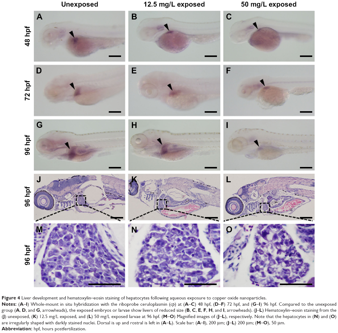
Liver development and hematoxylin–eosin staining of hepatocytes following aqueous exposure to copper oxide nanoparticles.
Notes: (A–I) Whole-mount in situ hybridization with the riboprobe ceruloplasmin (cp) at (A–C) 48 hpf, (D–F) 72 hpf, and (G–I) 96 hpf. Compared to the unexposed group (A, D, and G, arrowheads), the exposed embryos or larvae show livers of reduced size (B, C, E, F, H, and I, arrowheads). (J–L) Hematoxylin–eosin staining from the (J) unexposed, (K) 12.5 mg/L exposed, and (L) 50 mg/L exposed larvae at 96 hpf. (M–O) Magnified images of (J–L), respectively. Note that the hepatocytes in (N) and (O) are irregularly shaped with darkly stained nuclei. Dorsal is up and rostral is left in (A–L). Scale bar: (A–I), 200 μm; (J–L) 200 μm; (M–O), 50 μm.
Abbreviation: hpf, hours postfertilization.