
|
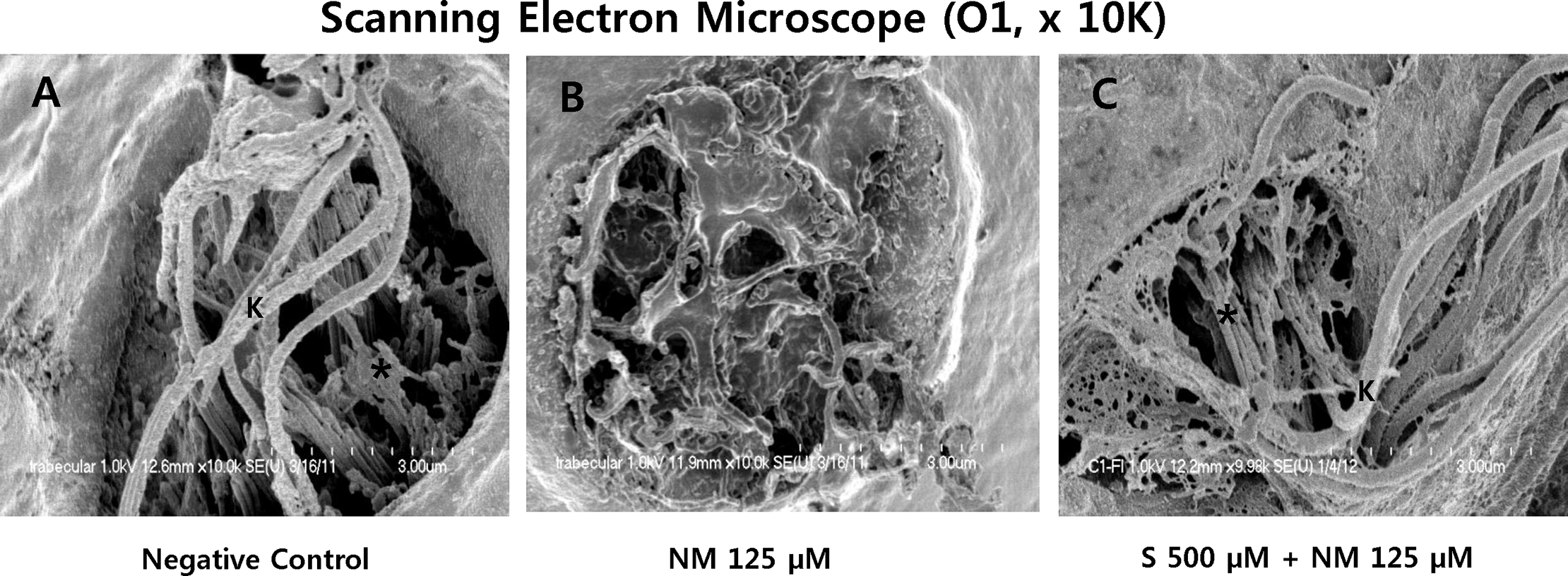
Fig. 5
Scanning electron microscopy (SEM, O1, x 10K).
The kinocilium (K) and the stereocilia bundles (asterisk) of hair cells in neuromast were clearly visible in the negative control (A). However, when the 5-dpf transgenic zebrafish were treated with 125μM neomycin for 1 h, the kinocilium and the stereocilia bundles were affected and severely disrupted (B). Sodium selenite provided nearly complete protection against neomycin-induced the damage of the kinocilium (K) and the stereocilia bundles (asterisk) in the neuromasts (C). Images were obtained in three 5-dpf zebrafish for each group. Scale bar (at the bottom of each figure, one space) = 3 μm. NM, neomycin; S, sodium selenite.