
Image
|
Figure Caption
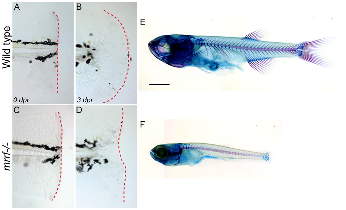
Fig. S2 Growth delay and defective fin regeneration in mrrf mutants. (A-D) Wild-type and mrrf-/- embryos had their tail fins amputated at 3 dpf. The dotted red lines show the caudal extent of the tail fin and the lack of regeneration in mutants 3 days post-resection (dpr). (E and F) Alcian blue and Alizarin red staining of cartilage and bone, respectively, show a severe growth defect in mrrf mutants at one month of age. Scale bar = 100 μm.
Figure Data
Acknowledgments
This image is the copyrighted work of the attributed author or publisher, and
ZFIN has permission only to display this image to its users.
Additional permissions should be obtained from the applicable author or publisher of the image.
Full text @ Development