
|
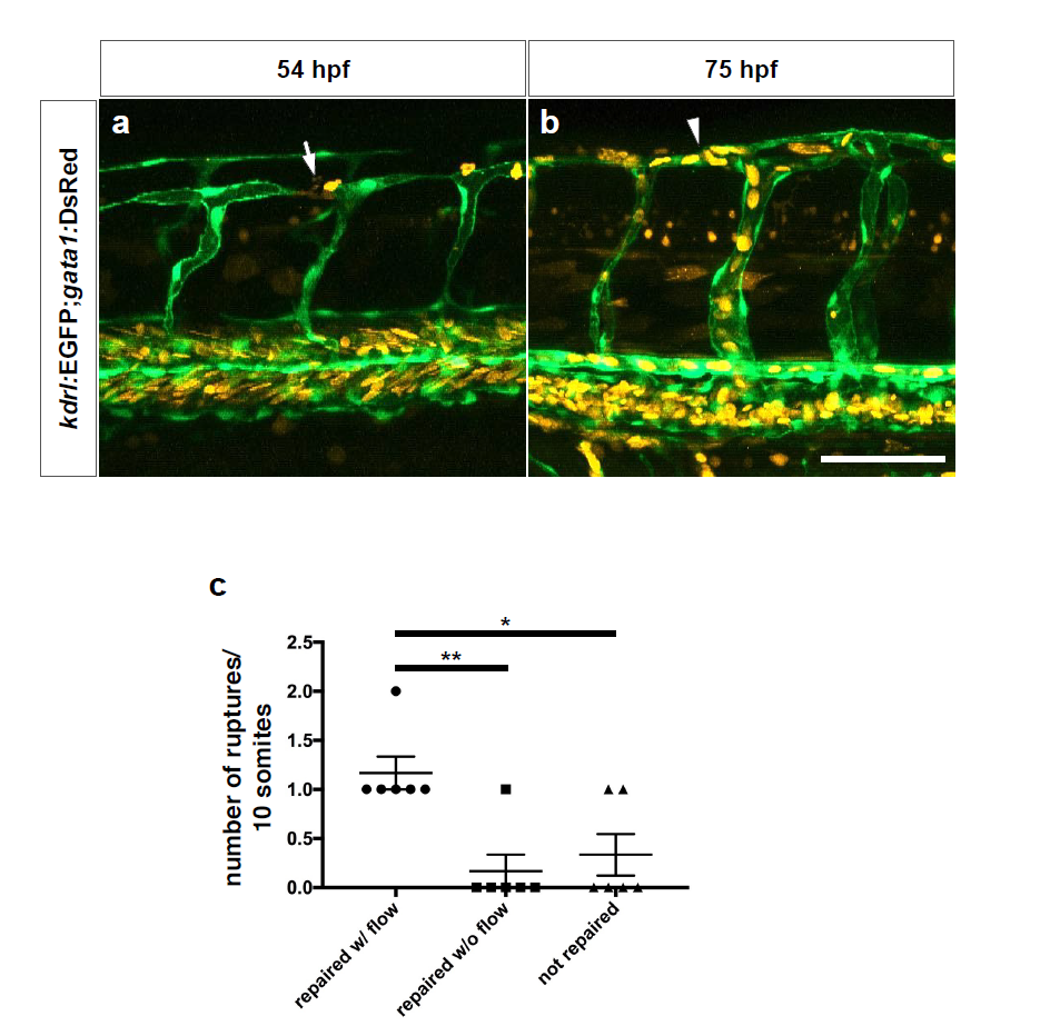
Fig. S10
Repaired blood vessels in WT embryos regain blood flow.
(a) Maximal intensity projections of confocal z-stacks of a blood vessel rupture in a 54 hpf Tg(kdrl:EGFP);Tg(gata1:DsRed) WT embryo after DMOG treatment for 6 hours starting at 48 hpf. (b) Maximal intensity projections of confocal z-stacks of the same blood vessel in the same embryo after repair. Lateral views; white arrow points to a ruptured vessel showing erythrocyte leakage, white arrowhead points to repaired vessel having regained flow. (c) Quantification of ruptured vessels subsequently repaired and showing presence (w/) or absence (w/o) of flow as well as those not repaired; data collected at 75 hpf. Bars represent mean ± s.e.m., n = 6 from 3 different clutches. (*P < 0.05; **P < 0.01; t-test). Scale bar, 50 μm.