
|
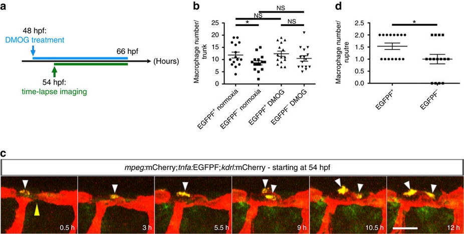
Fig. 8
Most macrophages closely associated with unstable vessels are tnfa:EGFPF+.
(a) Schematic representation of the experiment shown in b–d. (b) Quantification at 66 hpf of tnfa:EGFPF+ and tnfa:EGFPF− macrophages in normoxia and after DMOG treatment for 18 h starting at 48 hpf. (c) Maximal intensity projections of time-lapse confocal images of Tg(mpeg:mCherry);Tg(tnfa:EGFPF);Tg(kdrl:mCherry) WT embryo treated with DMOG starting at 48 hpf; lateral views. Yellow arrowhead points to an unstable vessel, white arrowheads point to recruited macrophages. (d) Quantification at 66 hpf of tnfa:EGFPF+ and tnfa:EGFPF− macrophages associated with 15 unstable vessels after DMOG treatment starting at 48 hpf. 38 macrophages were observed, 23 tnfa:EGFPF+ and 15 tnfa:EGFPF−. Bars represent mean±s.e.m., n=15 embryos from three different clutches, (*P<0.05; NS, no significant changes observed; t-test). Scale bar, 50 μm.