
|
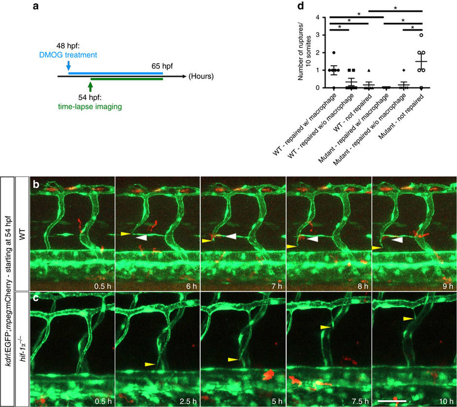
Fig. 7
Macrophages do not appear to assist in vessel repair in hif-1α mutants.
(a) Schematic representation of the experiment shown in b–d. (b) Maximal intensity projections of time-lapse confocal images of a Tg(kdrl:EGFP);Tg(mpeg:mCherry) WT sibling treated with DMOG starting at 48 hpf. Yellow arrowheads point to a vessel rupture and white arrowheads to macrophages. (c) Maximal intensity projections of time-lapse confocal images of a Tg(kdrl:EGFP);Tg(mpeg:mCherry) hif-1α mutant treated with DMOG starting at 48 hpf. Yellow arrowheads point to a vessel rupture and subsequent regression. (d) Quantification of blood vessel ruptures repaired in the presence (w/) or absence (w/o) of macrophages as well as those not repaired in WT and hif-1α−/− embryos at 65 hpf. All images represent lateral views. Bars represent mean±s.e.m., n=6 embryos from three different clutches, (*P<0.05; t-test). Scale bar, 50 μm.