
Fig. S1
- ID
- ZDB-IMAGE-170830-22
- Publication
- Xia et al., 2017 - Zebrafish slc30a10 deficiency revealed a novel compensatory mechanism of Atp2c1 in maintaining manganese homeostasis
- All Figures
- Figures for Xia et al., 2017
|
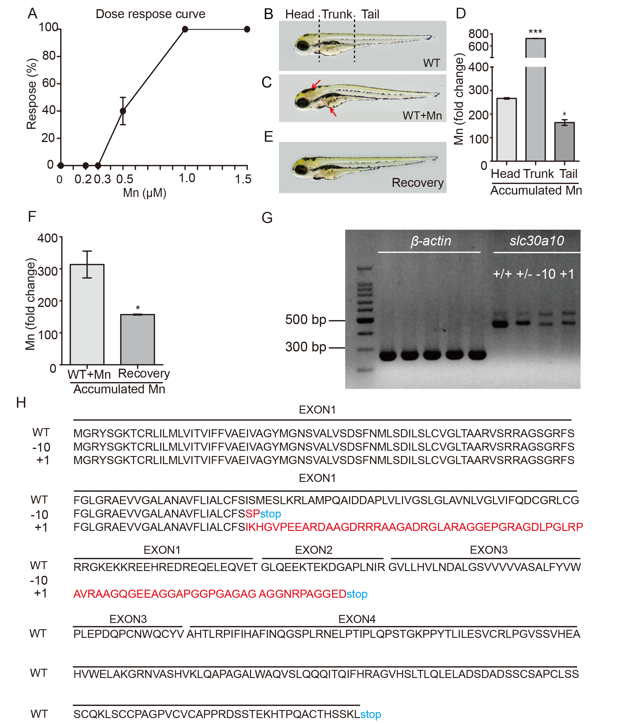
Fig. S1
Zebrafish embryos are a suitable model for studying Mn metabolism.
(A) Wild-type embryos were analyzed at 6 dpf following Mn exposure at the indicated concentration for 24 hours, and the percentage of embryos that responded (defined as impaired locomotion) is plotted against Mn concentration. (B and C) Mn accumulates in wild-type embryos exposed to 1 mM Mn for 24 hours, shown as a dark color in the brain and liver (C, red arrows). (D) Summary of the fold change in Mn in the three body regions of wild-type embryos exposed to 1 mM Mn for 24 hours. (E) 24 hours after transferring an Mn-exposed embryo to fresh Holt buffer, the color in the brain and liver returned to basal levels. (F) Summary of the fold change in Mn accumulation in Mn-exposed embryos that were transferred to fresh Holt buffer. (G) RT-PCR of slc30a10 mRNA in wild-type (+/+), heterozygous (+/-), homozygous 10-bp deletion (-10), and homozygous 1-bp insertion (+1) embryos. β-actin mRNA was measured as an internal control. (H) Predicted protein sequences of the wild-type (WT), -10, and +1 slc30a10 alleles generated using CRISPR/Cas9-based editing.