
|
Fig. 3
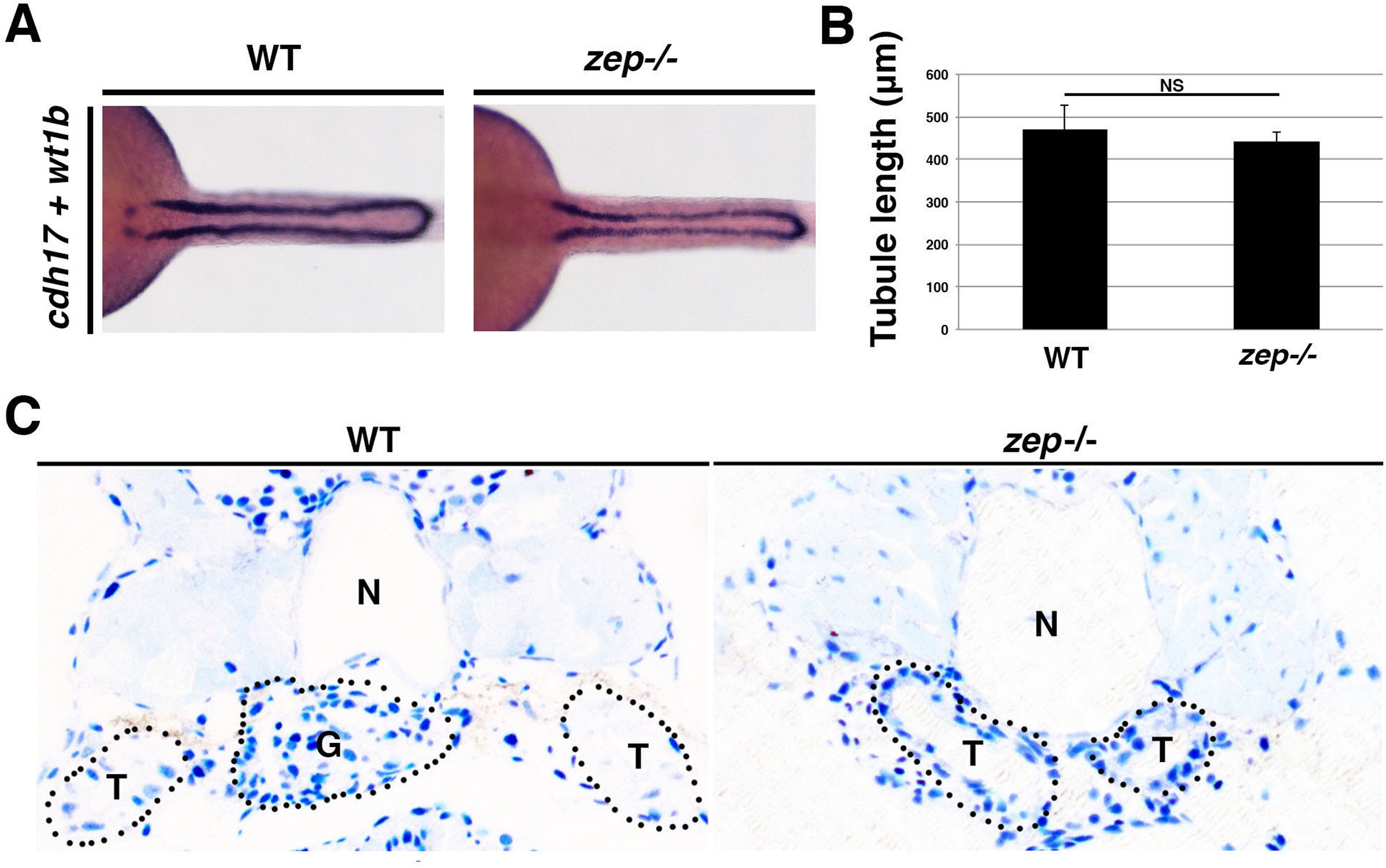
zepmutants show normal tubule development and fail to form a normal glomerulus. (A) WT and zep mutant embryos exhibit similar pairs of nephron tubules, which (B) are not statistically different in length. Embryos are shown in dorsal views. (C) Histological analysis of cross-sections (top, dorsal and bottom, ventral) from the cervical region of WT and zep embryos at 5 dpf. zep lack a midline glomerulus (G) ventral to the notochord (N), and the flanking tubules (T) are distended compared to WTs, consistent with the morphological appearance of edema. Black dotted lines outline the approximate perimeters of the indicated structures.
Reprinted from Developmental Biology, 428(1), Kroeger, P.T., Drummond, B.E., Miceli, R., McKernan, M., Gerlach, G.F., Marra, A.N., Fox, A., McCampbell, K.K., Leshchiner, I., Rodriguez-Mari, A., BreMiller, R., Thummel, R., Davidson, A.J., Postlethwait, J., Goessling, W., Wingert, R.A., The zebrafish kidney mutant zeppelin reveals that brca2/fancd1 is essential for pronephros development, 148-163, Copyright (2017) with permission from Elsevier. Full text @ Dev. Biol.