
Fig. 1
|
Fig. 1
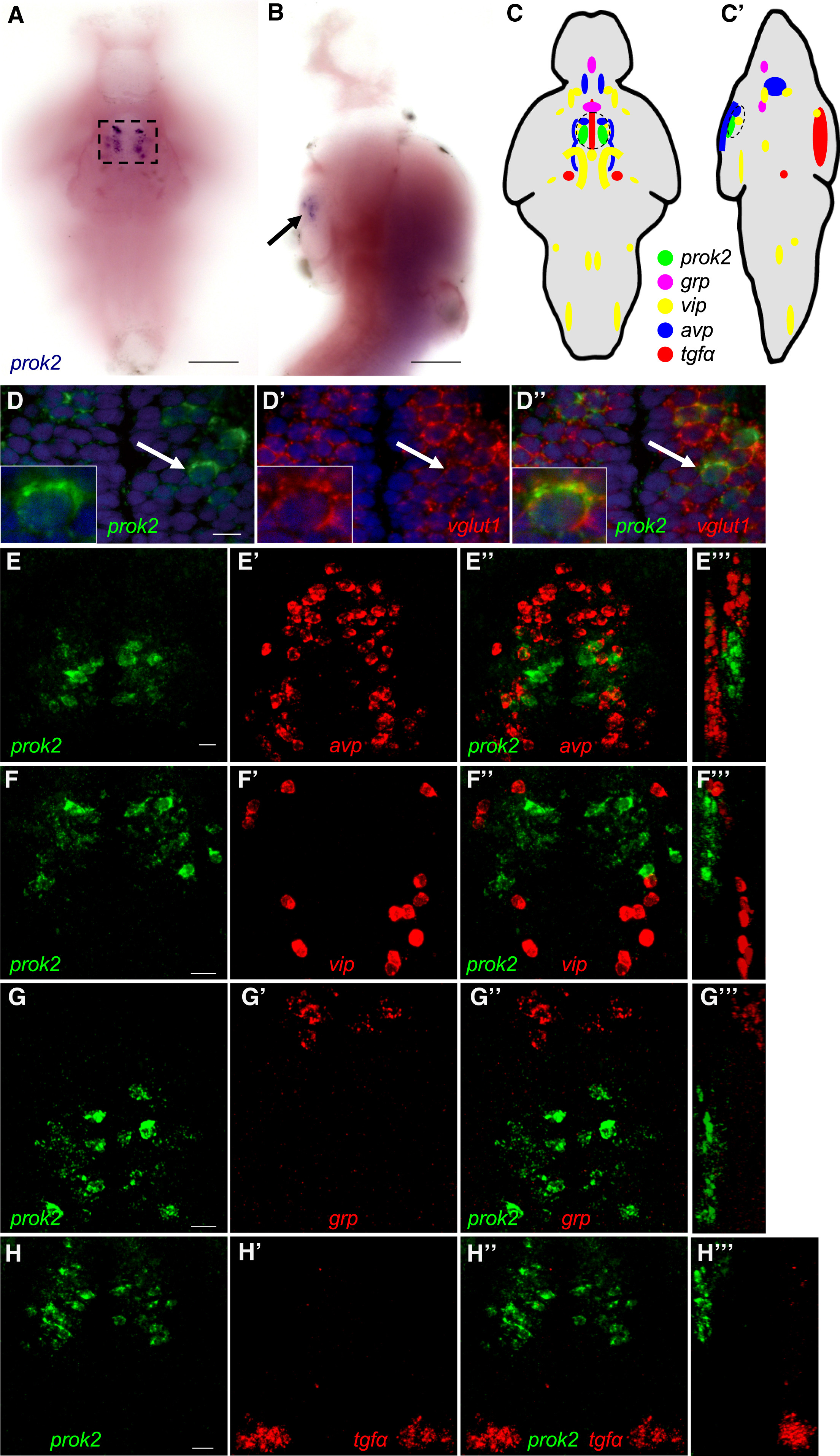
prok2 Expression in the Larval Zebrafish Brain
(A and B) prok2 mRNA (black box and arrow) is expressed in a bilateral cluster of approximately ten neurons in the ventral hypothalamus at 5 dpf. Dashed box in (A) indicates the region shown in (D). In (B), only the purple stain indicated by the arrow near the ventral brain surface is specific staining.
(C and C′) Schematic drawings summarize gene expression patterns from (E)–(H), as well as brain regions not shown in those panels. Dashed black circles indicate anatomical region potentially analogous to the mammalian SCN.
(D) prok2-expressing neurons co-express vglut1. DAPI nuclear stain is shown in blue. A representative cell, indicated by a white arrow, is enlarged in the inset. A 0.46-μm confocal section is shown.
(E–H) In the ventral hypothalamus, prok2 is expressed caudal and dorsal to populations that express avp (E) and caudal to cells that express vip (F). grp (G) and tgfα (H) are not expressed in the ventral hypothalamus. 34- (E–G) and 60- (H) μm-thick confocal projections are shown. (E′′′), (F′′′), (G′′′), and (H′′′) show orthogonal projections of each confocal stack. All samples are at 5 dpf. Ventral views with rostral at top (A, C–H, D′–H′, D″–H″) and lateral views with rostral at top and dorsal at right (B, C′, E′′′–H′′′) are shown. Scale bars in (A) and (B), 100 μm, and in (D)–(H), 10 μm.
See also Figure S1.