
|
Fig. S2
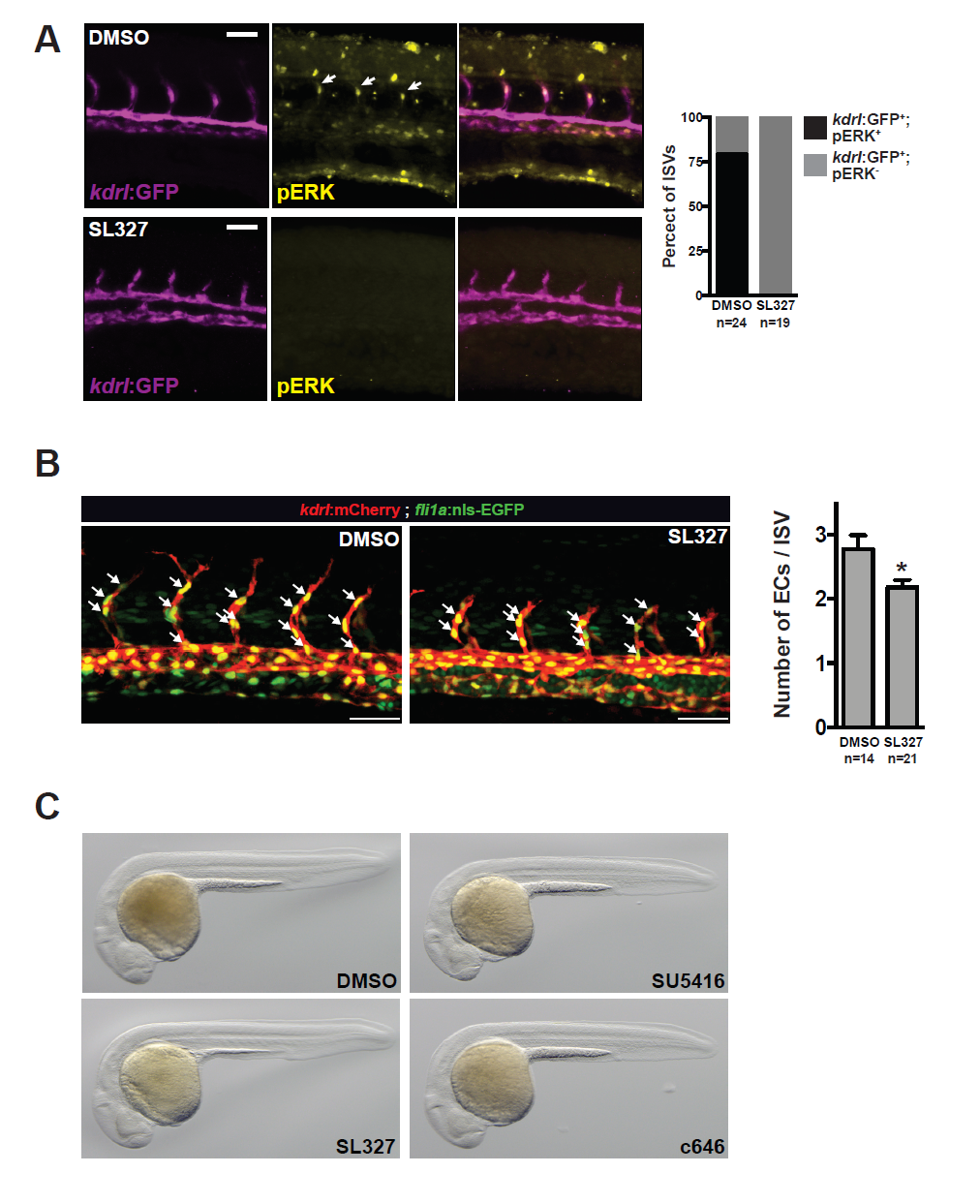
Characterization of sprouting defects in embryos treated with MEK inhibitor (SL327). A) pERK is enriched in tip cells during ISV formation in zebrafish and this is dependent on MEK signaling as it is negated in MEK-inhibitor (SL327) treated embryos (treated from 18-20 hpf to ~24 hpf). Quantification of the number of ISVs that stained positive for pERK is indicated. 24 hpf. Scale bar = 50 μm. B) The number of endothelial cells per intersomitic vessel was quantified in Tg(kdrl:mCherry;fli1a:nls-EGFP) embryos exposed to DMSO or SL327 (30 μM). The number of mCherry/EGFP double-positive cells was counted per intersomitic vessel and averaged per embryo. The number of embryos quantified is indicated. C) Transmitted light images of DMSO, SL327 (30 μM), SU5416 (5 μM) and c646 (3 μM) treated embryos revealing normal overall development and lack of tissue necrosis or developmental delay.