
|
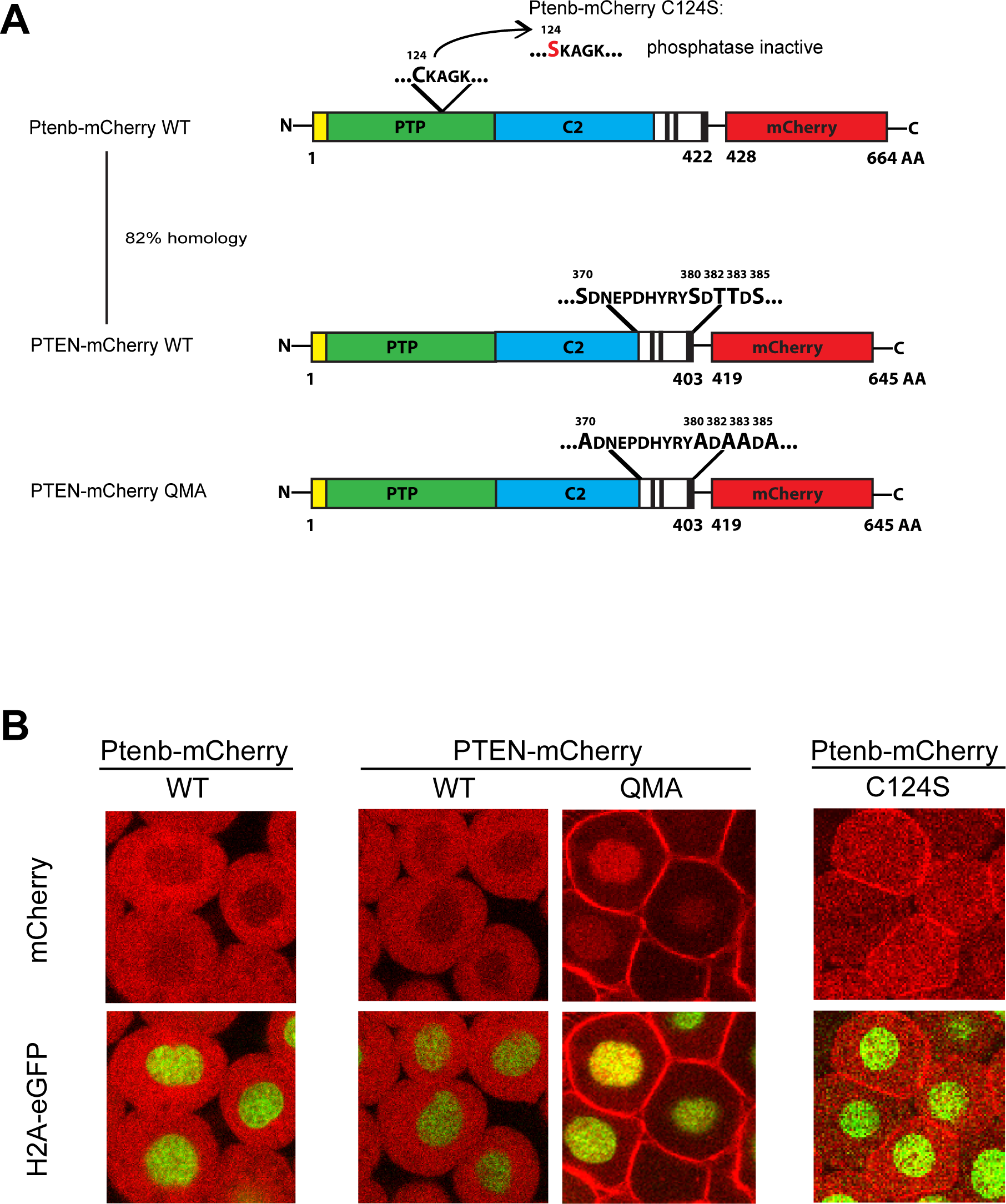
Fig. 1
PTEN QMA localizes to the cell membrane and nucleus in zebrafish embryos.
(A) Wild type Ptenb (long splicing variant, 422 amino acids) and human PTEN share a homology of 82%, both consisting of an N-Terminus (yellow), the PTP-domain (green), the C2 domain (blue) and the C-terminus (black and white). Mutation of the catalytic cysteine 124 to serine, C124S, results in catalytically inactive Ptenb. PTEN QMA contains five point mutations in its C-terminal phosphorylation sites, resulting in an open conformation of PTEN [19]. The red fluorescent protein mCherry (red) is C-terminally tagged in frame to all constructs used in these experiments. (B) Subcellular localization of Ptenb-mCherry, PTEN-mCherry, PTEN-mCherry QMA and Ptenb-mCherry C124S was assessed in tg(H2A-eGFP) zebrafish embryos, microinjected with 300pg synthetic mRNA at the one-cell stage and submitted to confocal live imaging at 6hpf. Top panels: mCherry expression; bottom panels: overlay of mCherry expression with H2A-GFP, which localizes to the nucleus.