
Fig. S1
- ID
- ZDB-IMAGE-170605-25
- Publication
- Zang et al., 2017 - Development of a Novel Zebrafish Model for Type 2 Diabetes Mellitus
- All Figures
- Figures for Zang et al., 2017
|
Fig. S1
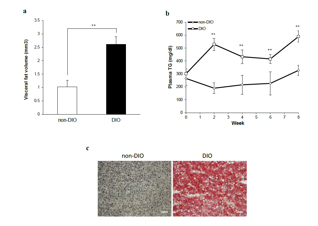
Assessment of visceral adipose tissue volume, plasma TG and hepatic steatosis in overfed zebrafish. (a) Changes in visceral adipose tissue volume in non-DIO and DIO zebrafish. Non-DIO group: n = 14; DIO group: n = 29. (b) Changes in plasma triglyceride (TG) levels in non-DIO and DIO zebrafish. Non-DIO group: n = 14; DIO group: n = 29. (c) Oil red O staining of liver sections. Red droplets indicate neutral lipid staining. Original magnification: × 400. Scale bar = 50 μm. Values are means ± SE. Statistical analyses were performed using Student’s t-test to compare the non-DIO and DIO groups at each time-point. *P < 0.05, **P < 0.01.