
|
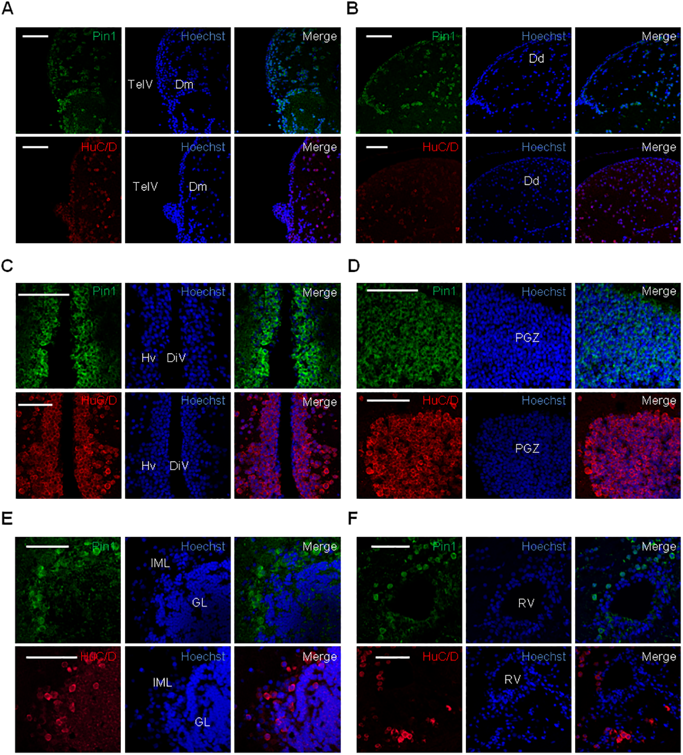
Fig. 6
Analysis of Pin1 expression in the adult zebrafish brain.
Immunofluorescence analysis on brain coronal sections using Pin1 (green) or HuC/D (red) as primary antibodies. (A) and (B) dorsal telencephalon, (C) ventral diencephalon, (D) optic tectum, (E) corpus cerebelli, (F) romboencephalic ventricular zone. DiV: diencephalic ventricle, Dd: lateral zone of dorsal telencephalon, Dm: medial zone of dorsal telencephalon, GL: cerebellar granular layer, Hv: ventral zone of periventricular hypothalamus, IML: cerebellar intermediate layer, PGZ: periventricular gray zone of the optic tectum, RV: romboencephalic ventricle, TelV: telencephalic ventricle. Scale bar = 50 μm.