
Image
|
Figure Caption
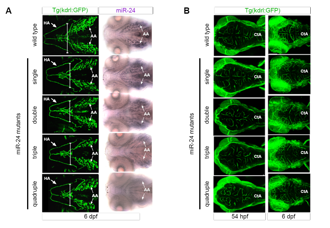
Fig. S4
miR-24Δ/Δ embryos do not have delay in development (related to Figure 4).
(A) Ventral view of Kdrl:GFP+ head vasculature (left) or mature miR-24 expression in the aortic arches (AA) (right). Mature miR-24 expression in AA diminishes upon increasingly loss of miR-24 gene copies. Arrows point to HA or AA.
(B) Dorsal head vasculature is similar between miR-24Δ/Δ and wild type genotypes. Cta = central arteries Developmental times as indicated.
Acknowledgments
This image is the copyrighted work of the attributed author or publisher, and
ZFIN has permission only to display this image to its users.
Additional permissions should be obtained from the applicable author or publisher of the image.
Reprinted from Developmental Cell, 40, Kasper, D.M., Moro, A., Ristori, E., Narayanan, A., Hill-Teran, G., Fleming, E., Moreno-Mateos, M., Vejnar, C.E., Zhang, J., Lee, D., Gu, M., Gerstein, M., Giraldez, A., Nicoli, S., MicroRNAs Establish Uniform Traits during the Architecture of Vertebrate Embryos, 552-565.e5, Copyright (2017) with permission from Elsevier. Full text @ Dev. Cell