
|
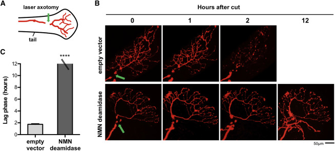
Fig. 1
NMN Deamidase Blocks Wallerian Degeneration in Zebrafish Larvae
(A) Schematic representation of larval zebrafish tail showing the location of the sensory (Rohon-Beard) axon and the site of injury (green arrow).
(B) One-cell-stage emrbyos were injected with plasmids for DsRed and NMN deamidase or empty vector. Time-lapse fluorescent images show DsRed-expressing axons in empty vector or NMN deamidase embryos after cut (green arrow shows the location of cut site). Axons have degenerated within 2 hr in the empty vector control, whereas NMN deamidase-expressing axons are preserved up to at least 12 hr.
(C) Length of the lag phase (time between the cut and the first sign of degeneration) in control or NMN deamidase-expressing axons (mean ± SEM; empty vector n = 9, NMN deamidase n = 8; Student’s t test, ∗∗∗∗p < 0.0001).