
Image
|
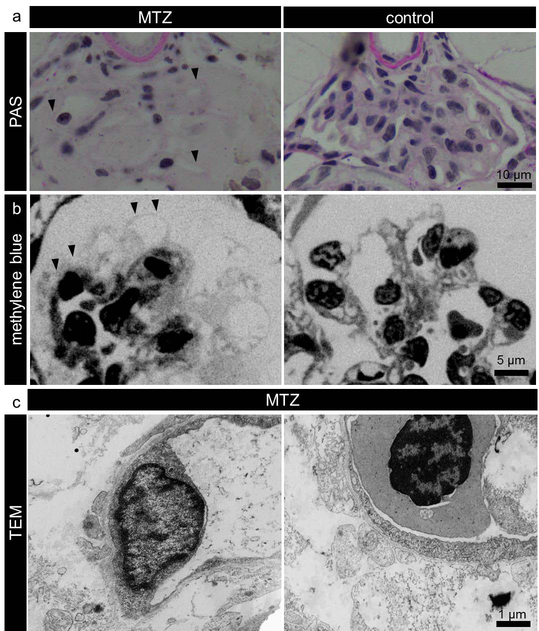
Figure Caption
Fig. S3
The denudation of the GBM. The PAS stained sections in panel a shows that broad areas of the glomerular capillaries were denuded and not covered by podocytes (a, arrowheads). Methylene blue stained semithin sections (panel b) showed absence of podocytes from the glomerular capillaries (b, arrowheads). Panel c shows additional transmission electron micrographs to Figure 2 b.
Acknowledgments
This image is the copyrighted work of the attributed author or publisher, and
ZFIN has permission only to display this image to its users.
Additional permissions should be obtained from the applicable author or publisher of the image.
Full text @ Sci. Rep.