
|
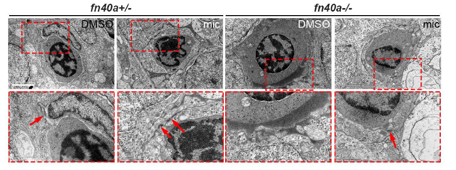
Fig. S4
Ultrastructures of microvessels in the zebrafish brain shown by transmission electron microscopy at 2.5 dpf (refer to Fig. 2). (A-D) Endothelial cells wraps well around red blood cells in fn40a heterozygotes with or without miconazole treatment (A-B). Endothelial cells outside red blood cells were disrupted in fn40a homozygote (C), which was rescued by miconazole treatment (D). RB, red blood cell; E, endothelial cell; Ne, neuron; mic, miconazole. Arrows point to the tight junctions between endothelial cells. The lower panels are magnification of the red-square area in the upper ones. Number of embryos per group was more than 8. Scale bar, 2 μm.