
|
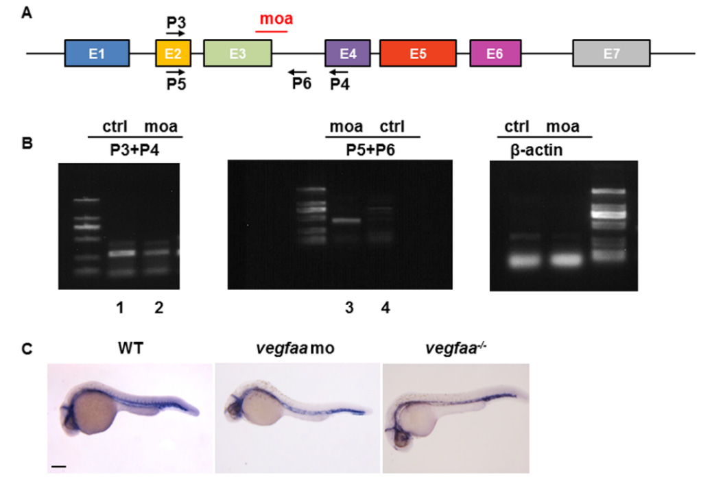
Fig. S2
RT-PCR validation of morpholino efficacy for vegfaa. (A) Open boxes and black lines denote exons and introns of the first coding and non-coding region of vegfaa. MO in red mark the target sites of the vegfaa translation blocking. P3, P4, P5, and P6 indicate the sites of primers; (B) One- to two-cell embryos were injected with vegfaa MO, and cDNA at 24 hpf was isolated. In wild-type embryos, the normal vegfaa spliced P3–P4 RT-PCR product was observed (lane 1) but not the P5–P6 splice donor site PCR product (lane 3). In vegfaa morphants, the normal transcrpiction level is decreased (lane 2) and the intron sequences are retained (lane 3). The β-actin cDNA was also amplified from each sample as a reference control; (C) Whole-mount in situ hybridizations (WISH) analysis of cdh5 in WT, vegfaa morphants, and mutants at 30 hpf. The morphants and mutants display identical phenotype in arterial-venous differentiation and intersegmental vessel sprouting. Scale bar: 200 μm.