
Fig. 6
|
Fig. 6
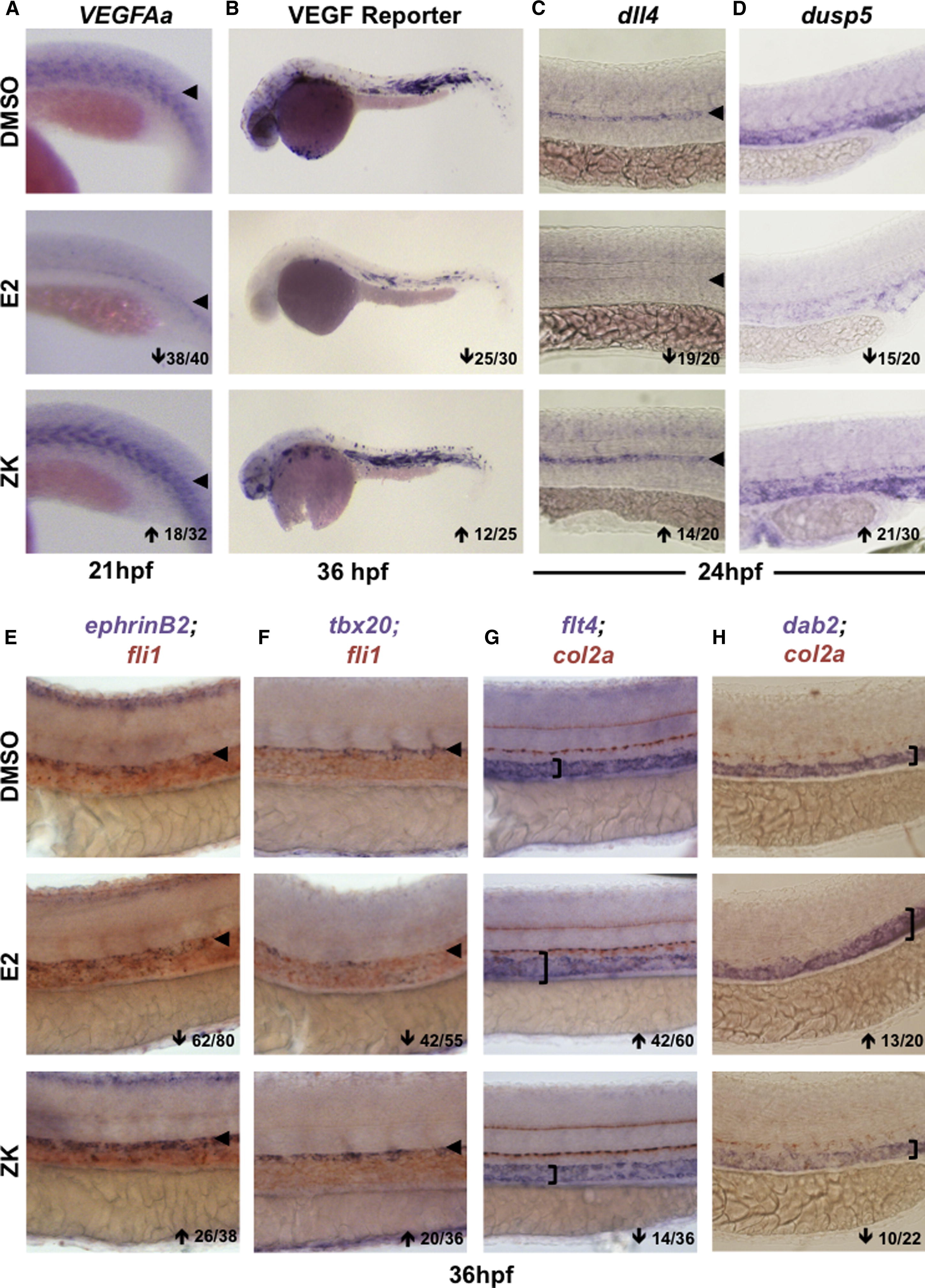
Blocking Esr Signaling Increases VEGF Activity and Alters Trunk Vasculature Patterning
(A) VEGFAa expression was enhanced after inhibition of endogenous estrogen activity via the pan-Esr antagonist ZK164015 (18/32).
(B) gfp expression in VEGF:GFP reporter embryos was reduced by E2 and enhanced by ZK (n > 25).
(C) The VEGF target gene dll4 was decreased after E2 exposure but increased by ZK (n > 20).
(D) dusp5 levels were reduced by E2 treatment but elevated after Esr antagonism (n > 20).
(E) Arterial ephrinB2 expression in the fli1+ vasculature was enhanced after ZK-mediated inhibition of estrogen signaling (26/38).
(F) tbx20 expression in the roof of the aorta was substantially increased by ZK (20/36).
(G) Venous flt4 was expanded dorsally toward the col2a+ hypocord by E2 but reduced in expression by ZK exposure (14/36).
(H) The venous marker dab2 was similarly regulated as determined by WISH (n > 20).