
|
Fig. S4
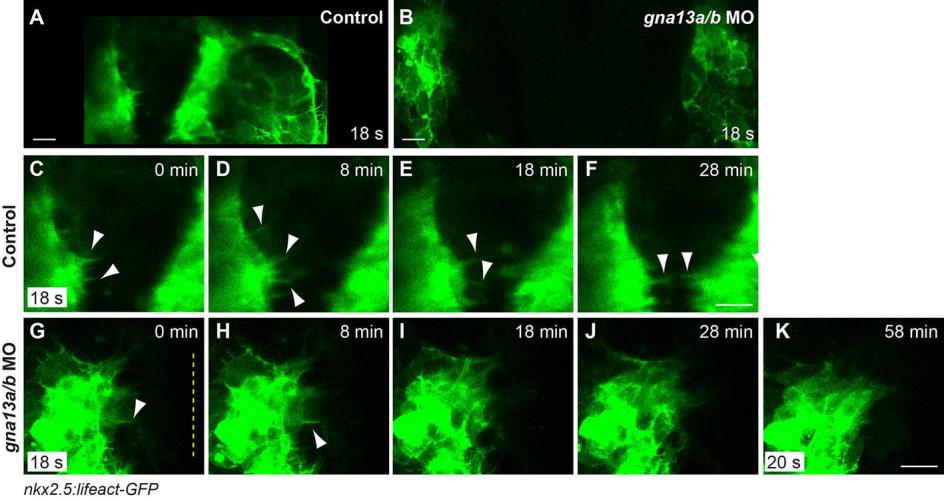
Myocardial precursors form robust migratory protrusions, which are transient in Gα13-deficient embryos. Snapshots from confocal movies of control and gna13a/b MO-injected Tg(nkx2.5:lifeact-GFP) embryos (supplementary movie 3). (A,B) The myocardial cells at 18s when the time-lapse started. (C-K) Magnified images in A-B at the indicated time-points. (C-F) The most dorsal regions of two myocardial populations in control embryos. (G-K) The left side of myocardial populations in morphants. The protrusions were presented initially, but disappeared in morphants. White arrowheads: protrusions. Yellow dashed line: midline. Scale bars: 20 μm.