
|
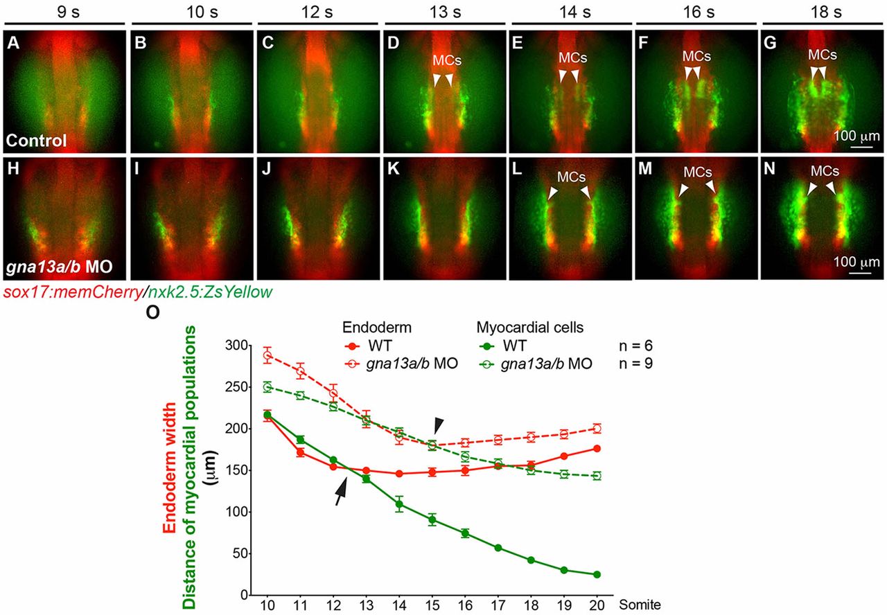
Fig. 4
Gα13 is required for all stages of myocardial migration. Epifluorescence time-lapse experiments performed on the embryos indicated (supplementary material Movie 2). (A-N) Snapshots of the anterior endoderm and myocardial cells from the movies at the stages indicated. Dorso-anterior views. White arrowheads indicate myocardial cells (MCs). (O) Endoderm width (red) and the distance between the two populations of myocardial cells (green) at the stages indicated. Black arrow and arrowhead denote the timepoints at which myocardial precursors were dissociated from the endoderm in the control and gna13a/b MO-injected embryos, respectively. Data are mean±s.e.m.