
|
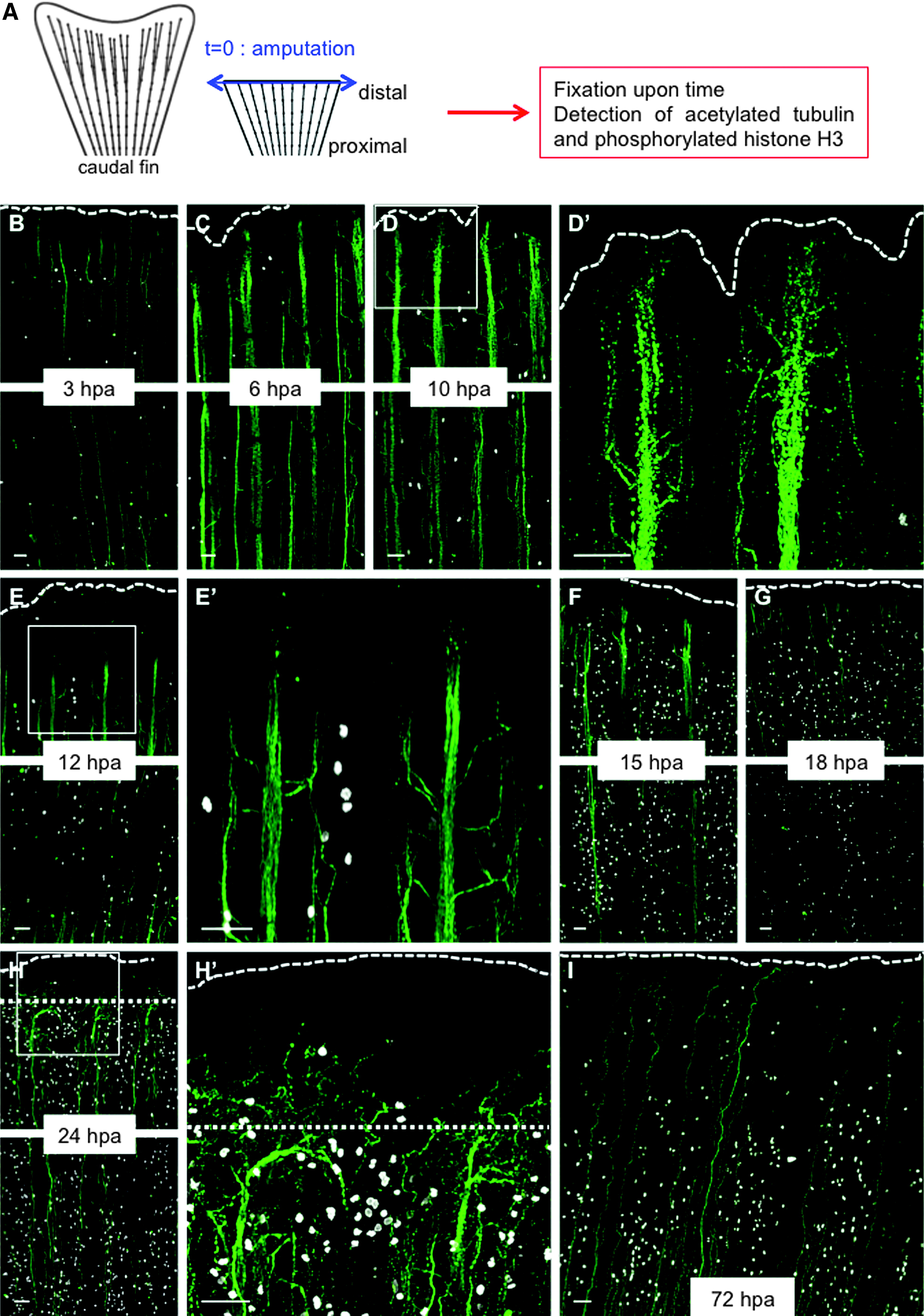
Fig. 1
Wallerian degeneration and axon growth after amputation are very rapid in adult fish. (A) Scheme of the experiment. (B–I) Caudal fins of adult fish were amputated (t = 0, blue arrow), and the axon cytoskeleton (green) and mitosis (white) were visualized over time through immunofluorescence staining for the axonal marker, acetylated tubulin (green), and phosphorylated histone H3 (white), respectively. (D′, E′, H′) Show higher magnifications of the distal parts of (D, E, H), respectively. At 6 hpa, the extremities of the axons begin to be fragmented, and the nerves begin to defasciculate; at 10 hpa, defasciculation is more pronounced. During Wallerian degeneration (10–12 hpa), epidermal and SCs start to proliferate. At 15 hpa, axons have regrown, and some axons cross the amputation plane at 18 hpa. Dotted line: amputation plane. Dashed line: distal part of the fin. Before 24 hpa, the amputation plane corresponds to the distal part of the fin. For each time point, the most distal part of the fin (upper panel) and a proximal part (lower panel) are shown. Scale bars = 50 μM. hpa, hours postamputation; SCs, Schwann cells.