
Image
|
Figure Caption
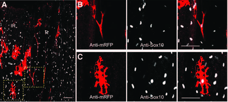
Fig. S4
After amputation, Sox10 is detected in neural crest-derived cells (melanocytes and SCs). The caudal fin of a pigmented sox10:mRFP transgenic fish was amputated, and the samples were fixed at 0.5 hpa. (A) Immunofluorescence staining for RFP (sox10:mRFP) (red) and Sox10 (anti-Sox10) (white) was performed. (B, C) Magnifications of (A). (B) Colocalization of mRFP and Sox10 in an SC. (C) Colocalization of mRFP and Sox10 in a melanocyte. Confocal images 1–2 µM; scale bar = 50 µM.
Acknowledgments
This image is the copyrighted work of the attributed author or publisher, and
ZFIN has permission only to display this image to its users.
Additional permissions should be obtained from the applicable author or publisher of the image.
Full text @ Antioxid. Redox Signal.