
|
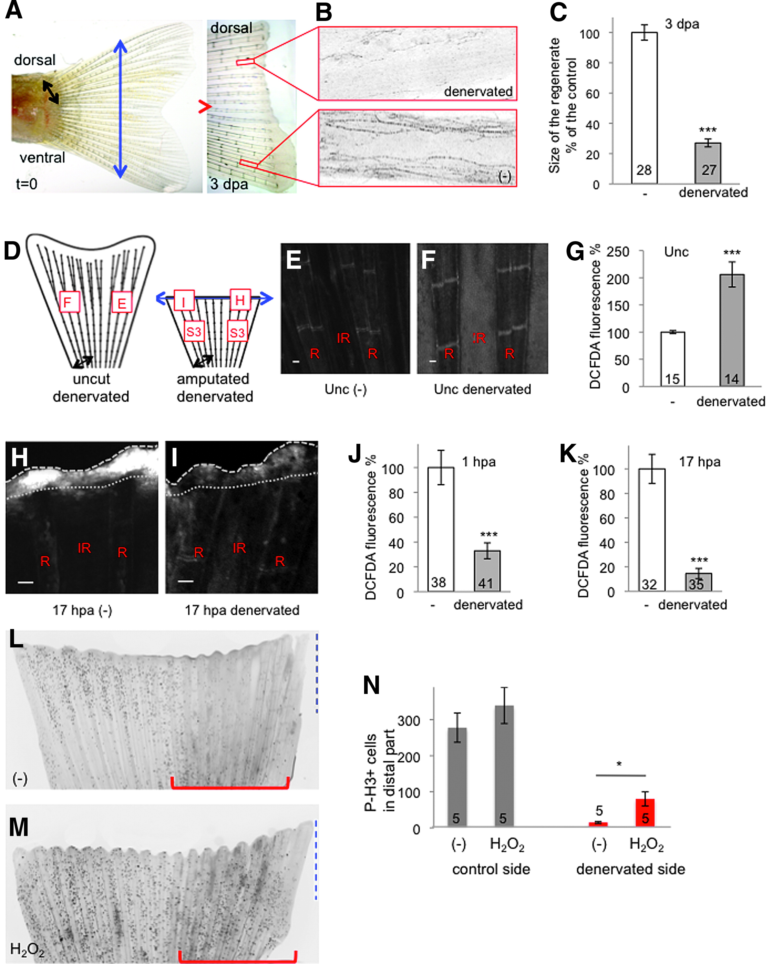
Fig. 3
Sensory neurons control ROS levels in the adult caudal fin. (A) The dorsal part of the caudal fin was denervated (black arrow) at the time of amputation (blue arrow). At 3 dpa, the dorsal part (denervated) had not regenerated compared with the ventral part (-) (representative image). (B) At 3 dpa, antiacetylated tubulin staining indicates the absence of axons in the denervated part. (C) Quantification of the size of the regenerated tissue at 3 dpa in the control (-) and denervated parts. The efficiency of regeneration is expressed as a percentage of the control. (D) Schematic representation of ROS detection in fin denervated in the dorsal part, with and without amputation. The squares indicate the position of ROS measurement, and the letters refer to panels in this figure or Supplementary Figure S3. (E–G) ROS detection at the level of the first ray bifurcation in an uncut fin. (H–K) ROS detection at the level of the amputation plane at 1 and 17 hpa. (H, I) Representative images at 17 hpa. (L–N) Mitotic cells were stained with antiphosphorylated histone H3 at 24 hpa in fins denervated at the time of amputation on the dorsal part (red line) and incubated in water (L) or 1 mM (H2O2) (M). Quantification of proliferation was performed on the distal part of the fin (blue dashed line) in the control part or the denervated part (red line) (N). The red line indicates the denervated part. (H, I) Dotted line: amputation plane. Dashed line: distal part of the fin. Error bars represent the SEM (*p < 0.05; ***p < 0.001). n Values are indicated at the bottom of each column of the graphs. Scale bars = 50 μM. A scheme of the different conditions in which ROS were measured is given in Supplementary Figure 2. dpa, days post amputation; H2O2, hydrogen peroxide; ROS, reactive oxygen species; SEM, standard error of the mean.