
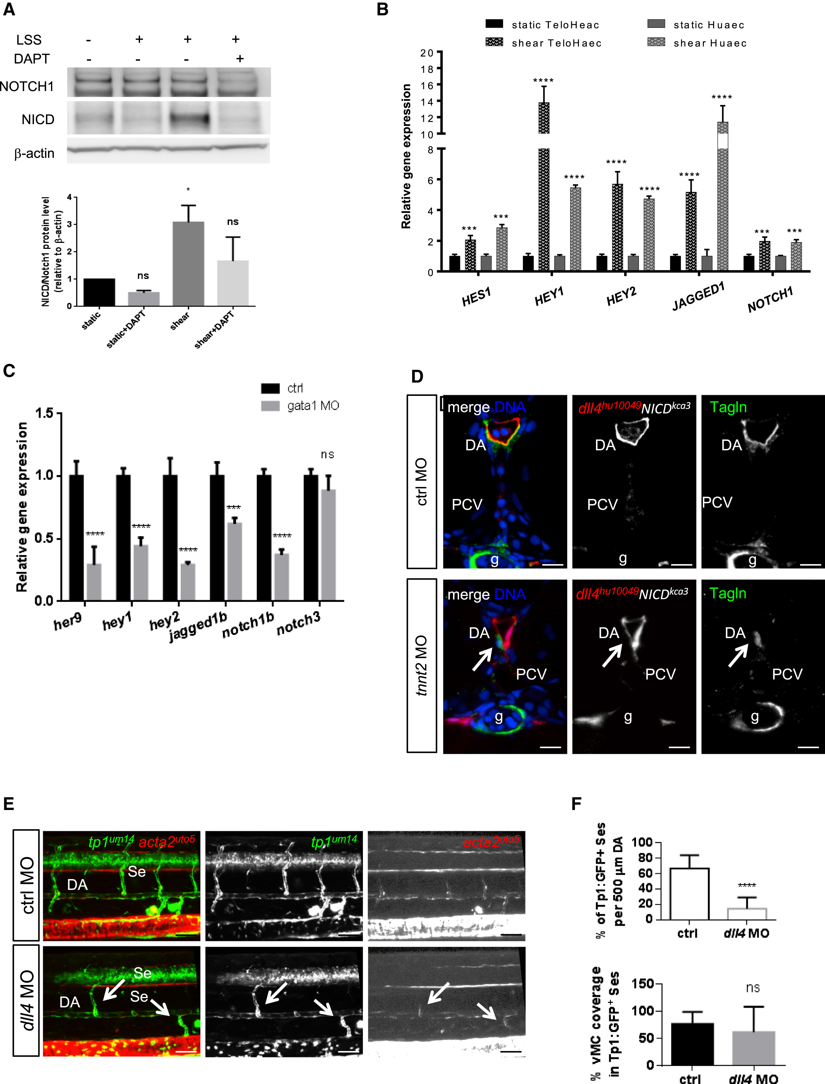
Fig. 4
|
Fig. 4
Notch Signaling Acts Downstream of Blood Flow to Recruit vMCs
(A) Shear-stress-mediated Notch activation in arterial-fated endothelial cells. Western blot analyses show increased levels of cleaved intracellular Notch (NICD) after 4 hr of LSS, whereas total Notch1 is unchanged. β-actin has been used as loading control. Histograms show the quantification of the amount of cleaved Notch1 (NICD)/over total Notch1 upon shear stress in endothelial cells.
(B) Shear stress induces Notch signaling in ECs. Effects of shear stress on the expression of different Notch target genes in human endothelial cell lines after 24 hr of LSS. ∗∗∗p < 0.001, ∗∗∗∗p < 0.0001. Data are represented as mean ± SD. Stars represent the results of two-way ANOVA.
(C) Loss of Notch signaling by blockade of blood flow in living zebrafish vessels. qPCR analyses of different Notch target genes are downregulated in ECs sorted from Tg(kdrl:egfp)s843 injected with control or gata1 MO at 48 hpf. ∗∗∗p < 0.001; ∗∗∗∗p < 0.0001. Data are represented as mean ± SD. Stars represent the results of two-way ANOVA.
(D) Arterial Notch activation restores vMC recruitment in the absence of blood flow in zebrafish vessels. Confocal images of Tagln staining (green) on section of TgBAC(dll4:GAL4FF;UAS:RFP)hu10049Tg(UAS:NICD)kca3 injected with tnnt2 or control morpholino. Expression of NICD in arterial ECs (dll4:RFP) rescue vMC recruitment (arrow) in the absence of blood flow (gata1 morphants). All the embryos analyzed (n = 15 in both conditions) show the indicated phenotype. Blue, nuclei. Scale bar, 25 μm.
(E) Blood flow can promote Notch signaling and vMC coverage in arterial vessels in the absence of dll4. Injection of dll4 morpholino induces a reduction of EGFP-positive Se vessels in Tg(tp1:egfp)um14 embryos. The few Se vessels that are positive for Notch activity and vMC coverage are the ones with circulation (arrow). A total number of 23 controls and 30 morphants were analyzed. Scale bar, 50 μm.
(F) Notch signaling is impaired after dll4 inactivation (KD). Histograms show the percentage of tp1:GFP+ Se vessels in 500 μm of DA, which are reduced in dll4 MO (n = 22/155 Se) compared to controls (n = 85/128 Se). The coverage of tp1+ vessels by vMCs is not altered in dll4 MO (n = 9/22) compared to controls (55/85), showing that flow can still recruit vMCs. A total number of 23 controls and 30 morphants were analyzed. Data are represented as mean ± SD. Stars represent the results of unpaired t tests of mean difference = 0 (∗p < 0.05, ∗∗p < 0.01, ∗∗∗p < 0.001).
Arrows indicate vessels with blood flow in dll4 morphants.
See also Figures S5 and S6.