
|
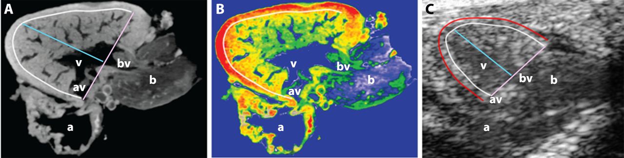
Fig. 2
Localization of anatomical landmarks in the zebrafish heart. (A) Representative optical section from a three-dimensional micro-computed tomography reconstruction of a wild-type, adult zebrafish heart (Skyscan 1072, ZEISS XRadia, Belgium). (B) False-colored heat map of the section shown in A demonstrating differing densities within the ventricular myocardium. The outer compact layer possesses greater density (red) in comparison with the highly trabeculated, non-compact spongy myocardium (yellow/green). (C) Representative high-frequency echocardiography B-mode image. In all three panels, the white border demarcates the interface between the compact and non-compact layers of the myocardium. The red border in C demarcates the outer layer of the epicardium. Pink line, basal diameter (DBASE), the distance between anterior and posterior borders of compact myocardium; blue line, perpendicular longitudinal diameter (DLONG) between ventricular base to apical compact myocardial border. a, atrium; av, atrioventricular valve; b, bulbus arteriosus; bv, bulboventricular valve; v, ventricle.