
|
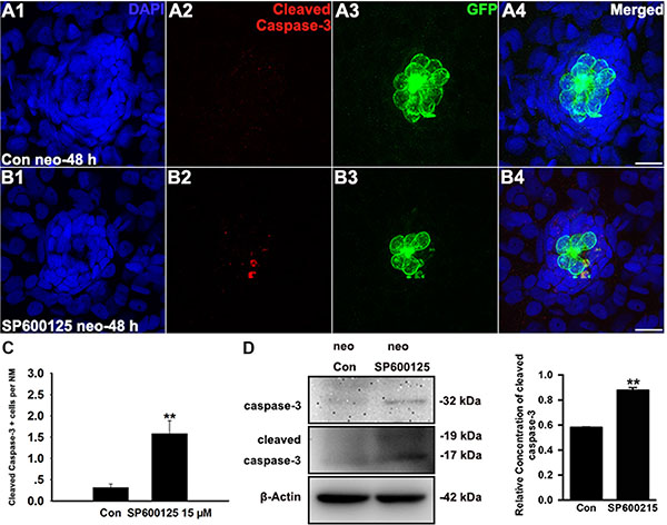
Fig. 2
SP600125 induces apoptosis in neuromasts. (A–B) Cleaved caspase-3 staining in the neuromast from a control larva (A) and 15 μM SP600125-treated larvae (B). Scale bar = 10 μm. (C) SP600125 treatment increased the numbers of cleaved caspase-3-positive cells. Bars are mean ± s.e.m. n = 48 control neuromasts and n = 48 15 μM SP600125-treated neuromasts. **p < 0.001. (unpaired t test, two-tailed, t = 4.051, df = 94, p < 0.001). (D) After treatment of larvae with 15 μM SP600125 for 48 h, protein extracts were prepared and subjected to western blot assay using antibodies against caspase-3 and cleaved caspase-3. β-Actin was included as the loading control.