
|
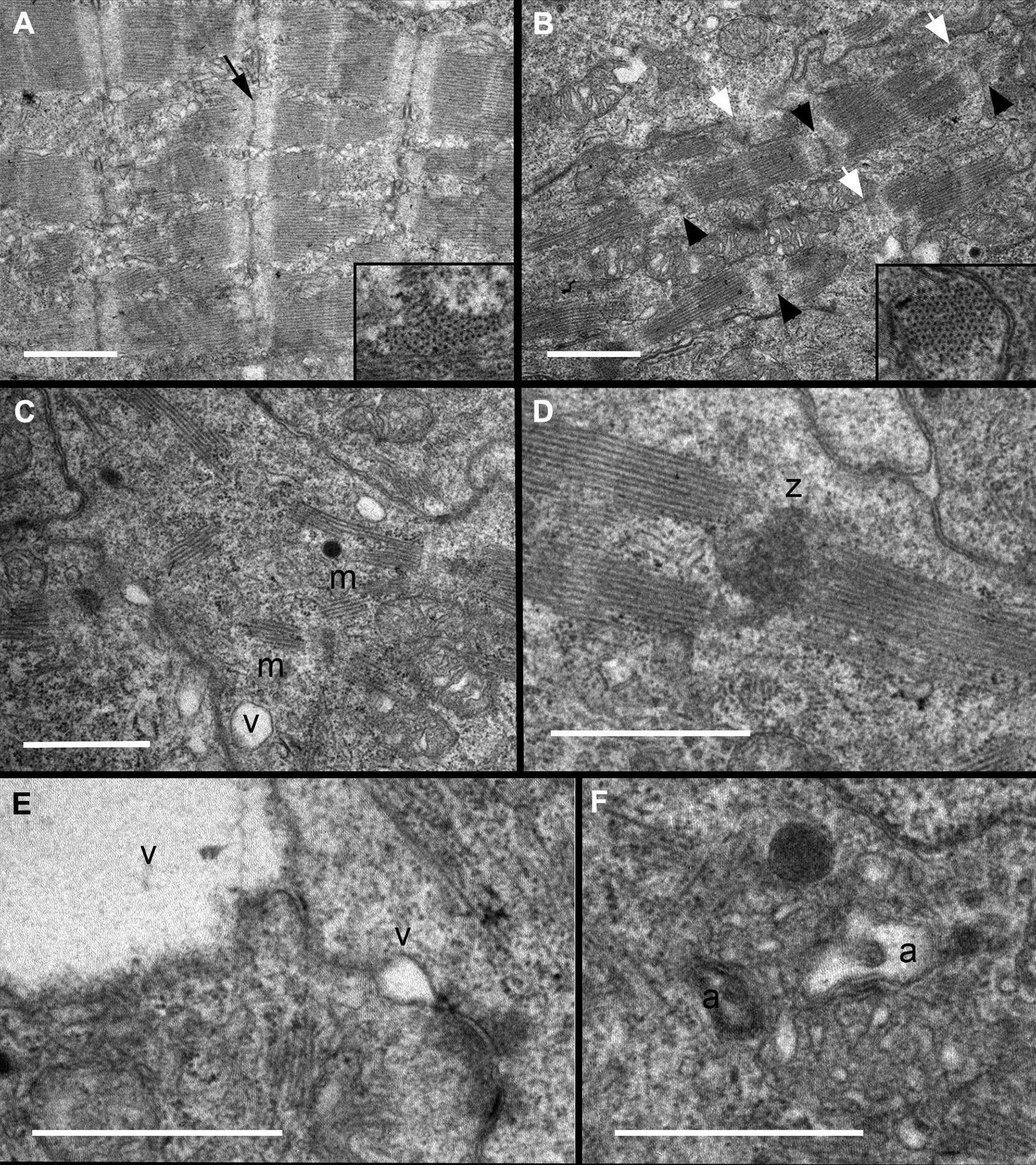
Fig. 6
ltrastructural Analysis Indicates Deficiencies in Sarcomere Organization and Z-Disc Formation
Transmission electron micrographs of zebrafish hearts at 72 hpf indicate that (A) wild-type embryos had assembled several consecutive sarcomeres with clearly distinct Z-discs (black arrow), whereas flncb+p53 MO hearts (B to F) show evidence of disorganized ultrastructure. (B) Most Z-discs were diffusely stained, irregular in shape (black arrowheads), or seemingly absent (white arrowheads). Insets: Cross section through myofilament bundles indicated a normal primary organization of thick and thin filaments in hexagonal lattices in flncb-depleted hearts. (C) Sarcomere arrangement in myofibrils (m) was often nonconsecutive, with (D) multiple sarcomeres sometimes adjoined to a mass of Z-disc-like material (z). (E) Small vacuoles (v) were often present between or near the plasma membranes of adjacent cardiomyocytes. These vacuoles could be large, creating a gap between adjacent cells. (F) Autophagic vesicles (a) were observed. Scale bar: 1 μmol/l. Abbreviations as in Figure 4.