
Fig. S7
- ID
- ZDB-IMAGE-170109-9
- Publication
- Takeuchi et al., 2015 - Type IV Collagen Controls the Axogenesis of Cerebellar Granule Cells by Regulating Basement Membrane Integrity in Zebrafish
- All Figures
- Figures for Takeuchi et al., 2015
|
Fig. S7
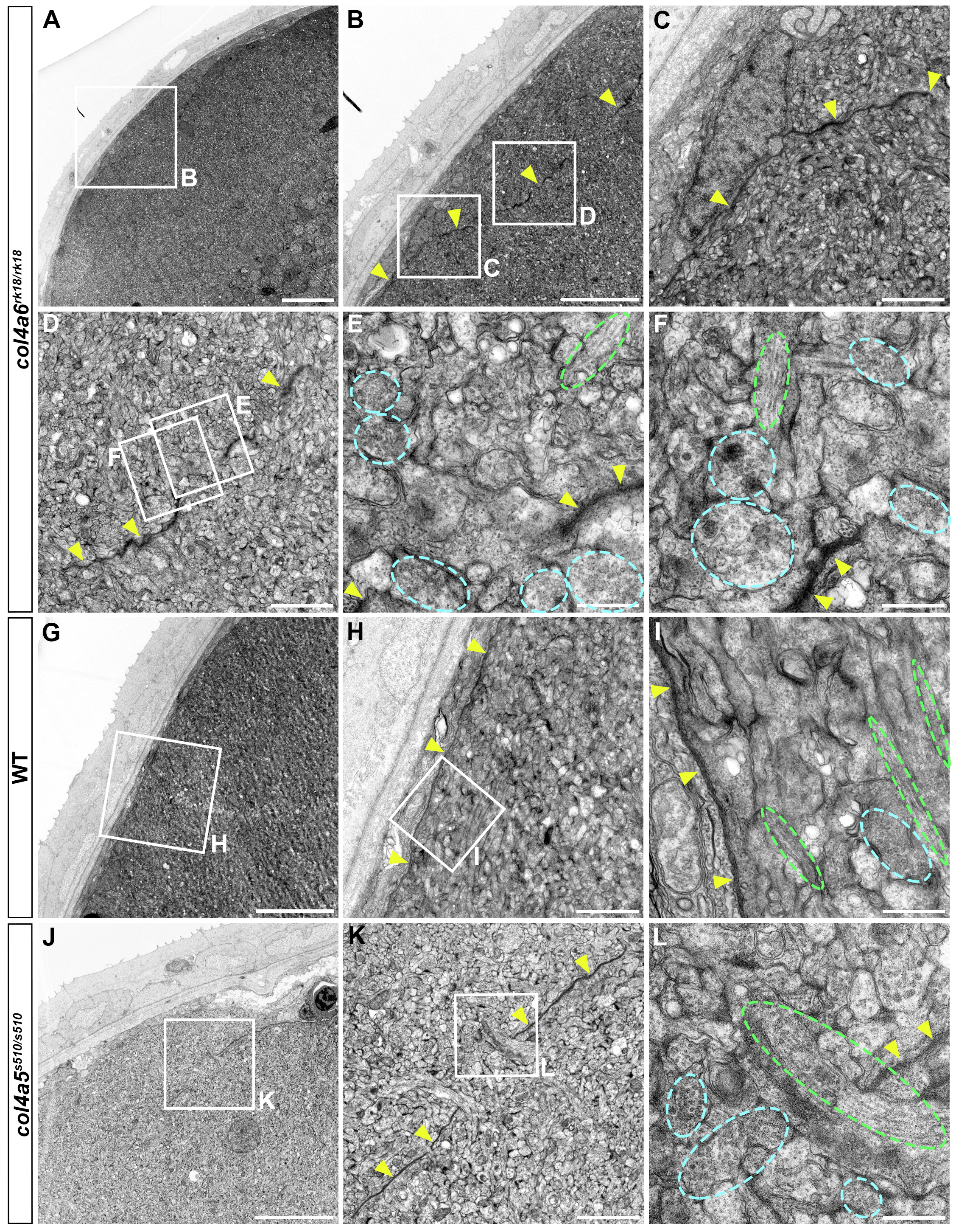
BM structure is disrupted in the col4a6 and col4a5 mutant tectum.
Tectum of 5-dpf wild-type (G-I), col4a6 mutant (A-F), and col4a5 mutant (J-L) larvae was analyzed by electron microscopy. Cross sections (A, G, J). (B) Higher magnification view of box B in (A). (C, D) Higher magnification views of boxes C and D in (B). (E, F) Higher magnification views of boxes E and F in (D). (H) Higher magnification view of box H in (G). (I) Higher magnification view of box I in (H). (K) Higher magnification view of box K in (J). (L) Higher magnification view of box L in (K). The BM is indicated by yellow arrowheads. Truncation of the tectal BM was observed in both col4a6 and col4a5 mutants. Axons containing synaptic vesicles and microtubules are marked by blue and green dashed circles, respectively. Scale bars: 20 μm in A; 10 μm in B, G and J; 2 μm in C, O, H and K; 0.5 μm in E, F, I and L.