
|
Fig. 6
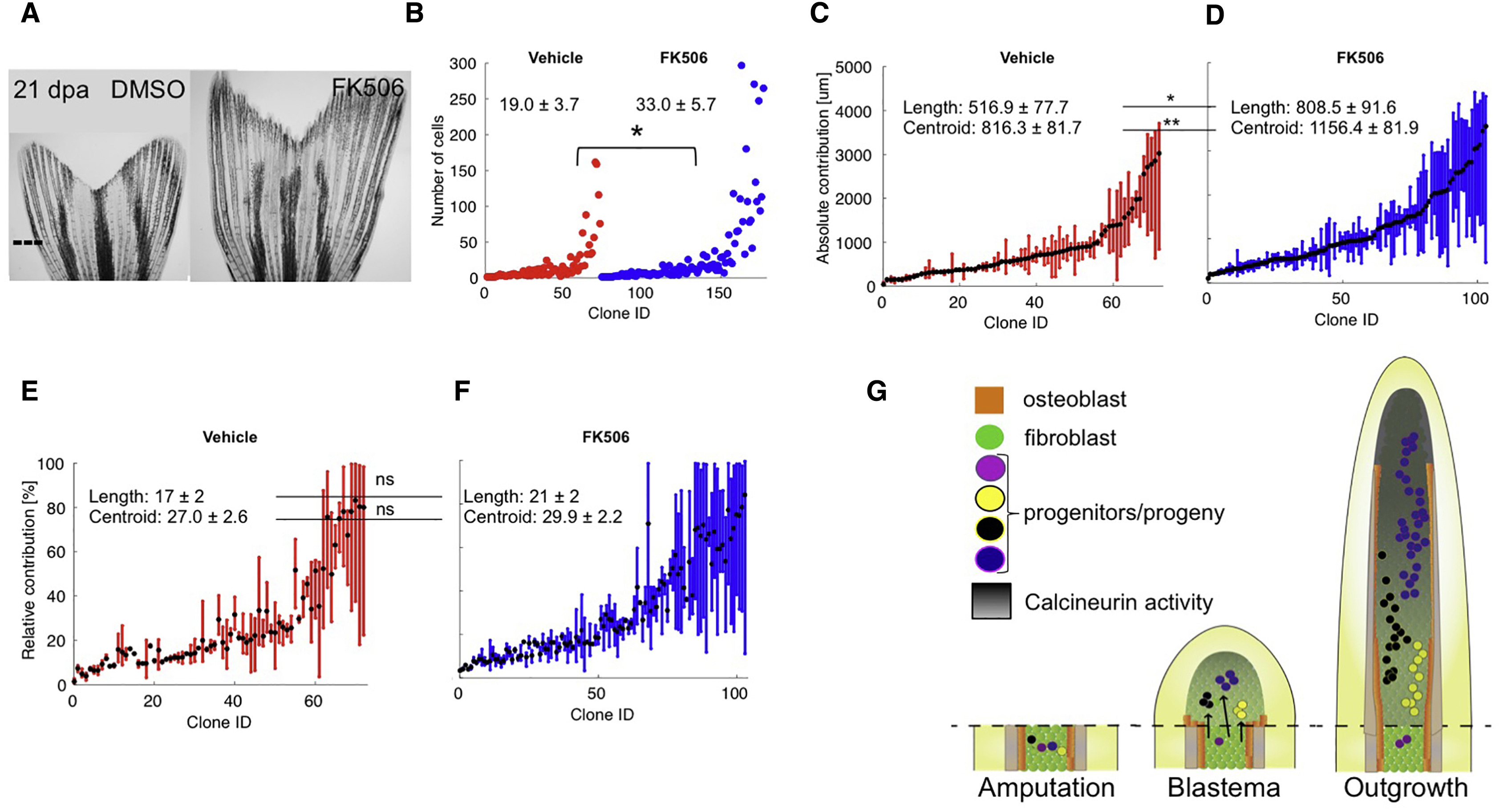
Calcineurin Enhances Regeneration by Scaling Clone Size
(A) Representative images of 21-dpa tph1b:CreER; ubi:switch fins treated with vehicle (DMSO) or FK506 from 3 to 21 dpa. Dashed line, amputation plane.
(B) Final numbers of cells in clones from vehicle- (red) or FK506-treated (blue) zebrafish at 21 dpa. ∗p < 0.05, Mann-Whitney one-tailed test, n = 73 (vehicle), n = 104 (FK506).
(C and D) Absolute positions along the PD axis (in μm) indicating clone contributions, for clones from vehicle- (C) or FK506-treated (D) fish. The centroid position of each clone is indicated with a black dot. Clone length is significantly increased in clones from FK506-treated fish, ∗p < 0.05, Mann-Whitney one-tailed test, n = 73 (vehicle), n = 104 (FK506). Centroid position is significantly distalized in clones from FK506-treated fish, ∗p < 0.005, Mann-Whitney one-tailed test, n = 73 (vehicle), n = 104 (FK506).
(E and F) Relative position of each clone, including the mean calculated centroid of clones from vehicle- (E) or FK506-treated (F) fish. Clone length and centroid positions are not significantly changed between the two groups, ns, not significant.
(G) Model for blastemal proliferation dynamics. Left: cells are recruited to form the blastema from regions proximal to the amputation plane. Middle: by 3 dpa, blastemal connective tissue progenitors establish preferences to contribute to distinct PD regions based on their coordinates. Right: the connective tissue compartment regenerates from progenitors in the blastemal approximate pre-pattern. Calcineurin activity increases as regeneration proceeds (dark gradient) and progressively lowers proliferation of fibroblast progeny. Blocking Calcineurin activity with FK506 releases the brake on cell division and clone cell number, scaling up the size of regenerated skeletal elements.
See also Figure S6.