
Fig. 6
- ID
- ZDB-IMAGE-161206-22
- Genes
- Publication
- Smith et al., 2014 - Contact-Mediated Inhibition Between Oligodendrocyte Progenitor Cells and Motor Exit Point Glia Establishes the Spinal Cord Transition Zone
- All Figures
- Figures for Smith et al., 2014
|
Fig. 6
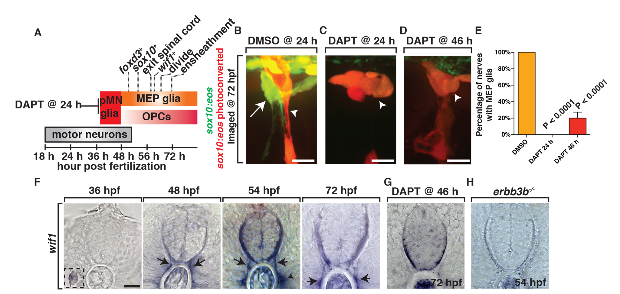
MEP glia originate from ventral spinal cord progenitors and express wif1.
(A) Summary of MEP glial development and the timing of marker expression. olig2 is not included because the initiation of expression is difficult to determine with surrounding olig2+ cells already present during this stage of development. (B–D) In Tg(sox10:eos) embryos exposed to UV light at 48 hpf and imaged at 72 hpf, (B) DMSO-treated animals have MEP glia, whereas (C) embryos treated with DAPT at 24 hpf and (D) 46 hpf do not. (E) Quantification of data from panel D. (F) In situ hybridization with wif1 riboprobe at 36, 48, 54, and 72 hpf shows timing of wif1 expression at the root denoted by arrows. Arrowhead denotes the horizontal myoseptum staining. Inset at 36 hpf shows wif1+ staining is expressed at the lateral line, as has been previously described. (G–H) At 54 hpf in erbb3b (H) mutants and at 72 hpf in embryos treated with DAPT at 46 hpf (G), we observed an absence of wif1 staining along spinal motor roots. Scale bars, 25 µm.