
Fig. 4
- ID
- ZDB-IMAGE-161206-20
- Genes
- Publication
- Smith et al., 2014 - Contact-Mediated Inhibition Between Oligodendrocyte Progenitor Cells and Motor Exit Point Glia Establishes the Spinal Cord Transition Zone
- All Figures
- Figures for Smith et al., 2014
|
Fig. 4
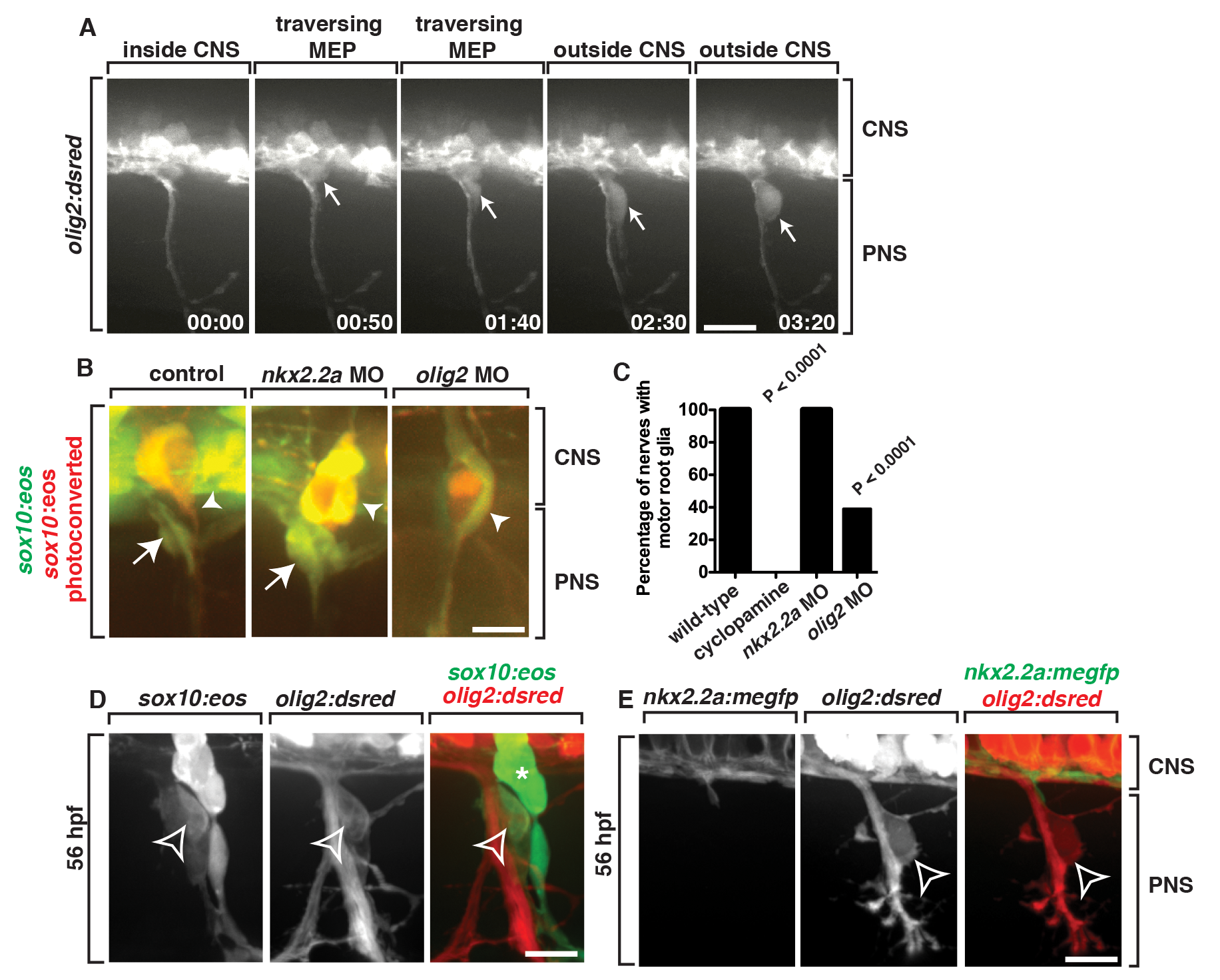
sox10+ motor root glia arise from olig2+ precursors in the spinal cord.
(A) Frames captured from a 24-h time-lapse video of a Tg(olig2:dsred) larvae showing olig2+ cells (arrow) migrating out of the CNS, pinching at the MEP, and associating with the spinal motor root axons. Numbers in lower right corners denote time lapsed from the first frame of the figure. Brackets show the CNS and PNS portions in the images. (B) In Tg(sox10:eos) embryos injected with a nkx2.2a MO, CNS-derived sox10+ motor glial cells (arrow) are indistinguishable from wild-type nerves. In contrast, in olig2 MO-injected embryos, motor root glial cells are absent. In both instances, sensory glial cells are unaffected (arrowheads). (C) Quantification of data from panel B (>50 nerves were scored for each perturbation). (D) In a Tg(sox10:eos);Tg(olig2:dsred) embryo at 56 hpf, a sox10+ cell located on the motor nerve root expresses olig2 and sox10 (open arrowhead). Neighboring sox10+ cells in the DRG (asterisk) do not express olig2. (E) In a Tg(nkx2.2a:megfp);Tg(olig2:dsred) embryo at 72 hpf, nkx2.2+ perineurial glia are distinct from olig2+ MEP glia (open arrowhead). All images are lateral views of the motor and sensory root with dorsal to the top and anterior to the left. Scale bars, 25 µm.