
IMAGE
Fig. 2
- ID
- ZDB-IMAGE-161206-18
- Genes
- Publication
- Smith et al., 2014 - Contact-Mediated Inhibition Between Oligodendrocyte Progenitor Cells and Motor Exit Point Glia Establishes the Spinal Cord Transition Zone
- All Figures
- Figures for Smith et al., 2014
Image
|
Figure Caption
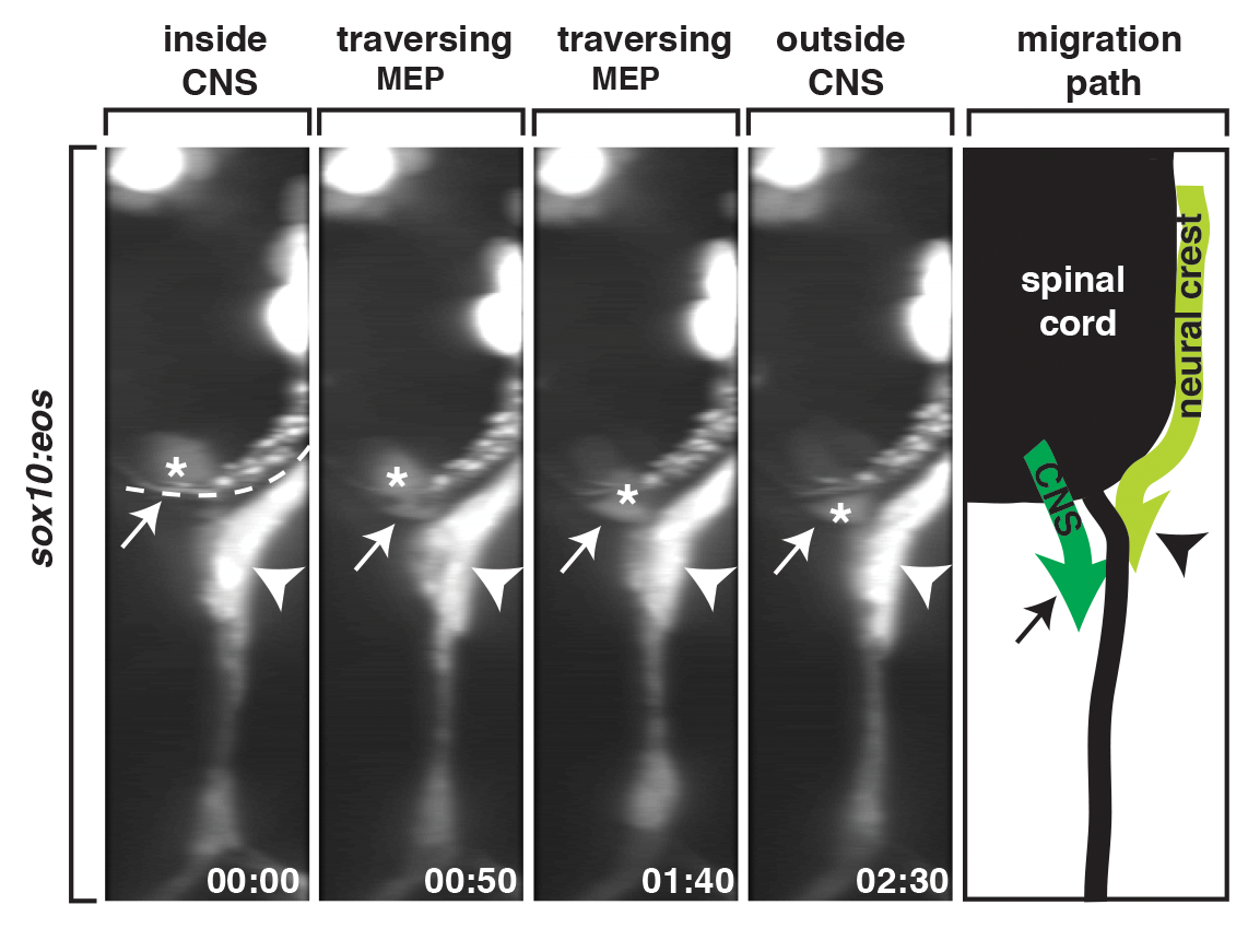
Fig. 2
sox10+ motor root glial cells originate in the CNS.
A 90-degree rotation of images captured from a 24-h time-lapse video beginning at 48 hpf in a Tg(sox10:eos) embryo. Numbers in lower right corners denote time lapsed from the first frame of the figure. At approximately 56 hpf (00:00), a sox10+ cell (asterisk) migrated from the spinal cord, pinched through the MEP (arrow), and migrated into the periphery. Migration path shows the migration of neural crest versus CNS-derived sox10+ cells during this developmental stage. Dashed line marks lateral edge of the spinal cord. Scale bar, 25 µm.
Figure Data
Acknowledgments
This image is the copyrighted work of the attributed author or publisher, and
ZFIN has permission only to display this image to its users.
Additional permissions should be obtained from the applicable author or publisher of the image.
Full text @ PLoS Biol.