
|
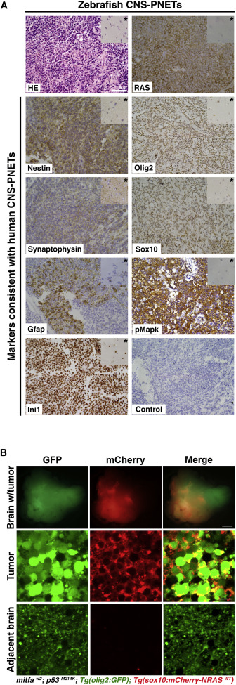
Fig. 2
Zebrafish NRASWT-Driven Brain Tumors Derived from the Anterior Brain Closely Resemble Oligoneural CNS-PNET Tumors
(A) Shown is a representative example of the histology (H&E [HE]) and IHC analysis (indicated in each panel) from three independent zebrafish brain tumors arising in the anterior lobes of NRASWT-expressing fish. Inset in each top right corner (indicated by a star) shows staining in the adjacent normal brain area. Scale bars, 50 μm.
(B) Top panels show a confocal image of a live stable transgenic [mitfaw2; p53M214K; Tg(olig2:GFP)] zebrafish brain with an optic tectum tumor expressing Tg(sox10:mCherry-NRASWT). Middle panels show that the tumor is composed of large undifferentiated cells expressing GFP in the cytoplasm and mCherry at the membrane. Bottom panels show typical elongated oligodendrocytes in the adjacent tumor-free area. Scale bars, 500 μm (brain with tumor) and 10 μm (tumor and adjacent brain).