
Image
|
Figure Caption
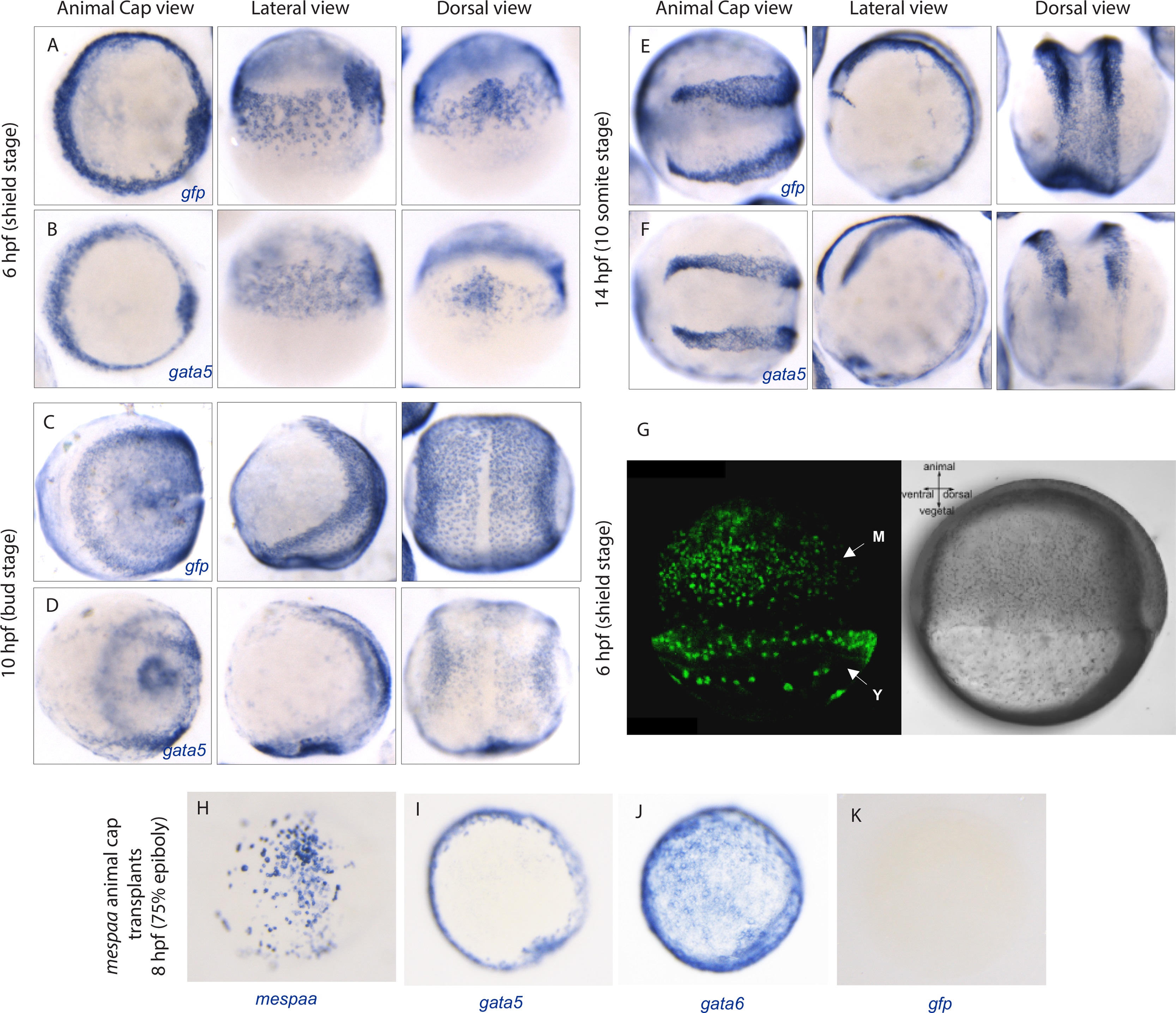
Fig. S3
gata5 and gfp are expressed in the same pattern in gata5:EGFP transgenic embryos. (A-F) gata5 and gfp expression in gata5:EGFP embryos at 6, 10 and 14 hours post fertilization. Embryos are viewed from the animal cap, lateral and dorsal sides. (G) gata5:EGFP transgenic embryo at 6 hpf (shield stage). M indicates cells at the embryonic margin while Y indicates the yolk syncytial layer (YSL). (H-K) mespaa, gata5, gata6 and gfp expression in animal cap transplants of mespaa over-expressing Tg(gata5:EGFP) cells in host embryos at 75% epiboly (8 hpf). Embryos are visualized from an animal cap view.
Figure Data
Acknowledgments
This image is the copyrighted work of the attributed author or publisher, and
ZFIN has permission only to display this image to its users.
Additional permissions should be obtained from the applicable author or publisher of the image.
Reprinted from Developmental Biology, 418(1), Deshwar, A.R., Onderisin, J.C., Aleksandrova, A., Yuan, X., Burrows, J.T., Scott, I.C., Mespaa can potently induce cardiac fates in zebrafish, 17-27, Copyright (2016) with permission from Elsevier. Full text @ Dev. Biol.