
|
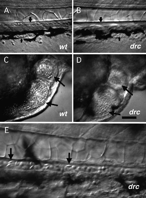
Fig. 6
Blood cells are photosensitive in drc mutants. (A-D) DIC videomicroscopy of 2.5 dpf embryos using low-light level illumination and a SIT camera with frame-averaging. All embryos were obtained from a single cross between two drcm87 heterozygotes. Circulation was stopped in the embryos by tricaine overdose. (A) The tail of a phenotypically wild-type embryo raised continuously in room light. Blood cells are seen in the caudal artery (large arrow) and caudal vein (small arrow). (B) The tail of a mutant sibling raised continuously in room light The artery and vein are both present (arrows), but contain no blood cells. (C) Blood cells in the heart of a phenotypically wild-type embryo raised in darkness from blastula stages on. (D) Blood cells in the heart of a mutant sibling embryo raised in darkness from blastula stages on. Abundant blood cells are seen in both the phenotypically wild-type and mutant embryos (arrows). (E) Higher magnification view of blood cells in the caudal artery of a mutant embryo. Blood cells with the typical lentiform morphology for this age are seen (arrows). Scale bar, 100 µm (A-D), 29 µm (E).