
|
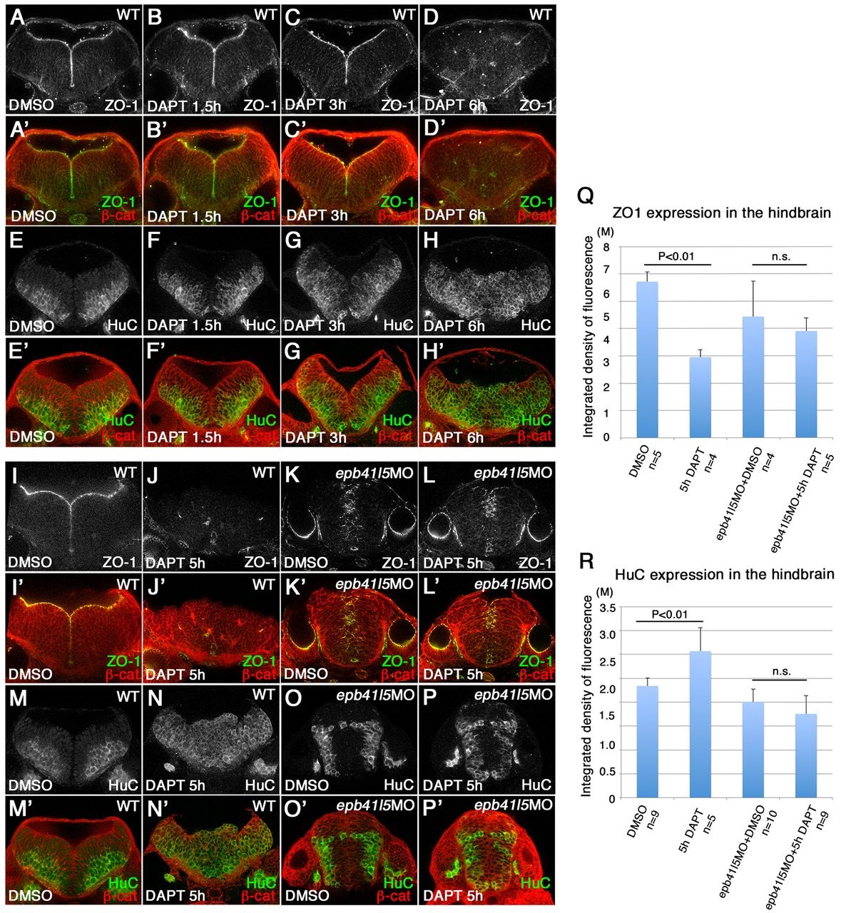
Fig. 3
Delays in disassembly of AJs and neuronal differentiation in epb41l5 morphants. (A-H′) Timeline of AJ disassembly (A-D′) and differentiation of neurons (E-H2) during DAPT treatment in wild-type embryos. Embryos were treated with 50µM DAPT for 1.5, 3 or 6h and immunostained at 32hpf. Loss of Notch signaling by DAPT treatment results in loss of AJs and premature differentiation of NPCs into neurons. (I-P′) Delays in AJ disassembly and differentiation of neurons in epb41l5 embryos. Embryos were treated with DAPT for 5h. In control embryos (WT), 5h DAPT treatment eliminates ZO-1 at the apical/ventricular surface, accompanied by a corresponding increase in HuC. In epb41l5 morphants, there is a much smaller reduction in ZO-1 immunostaining and no obvious increase in HuC immunostaining. (Q,R) Quantitative analyses of ZO-1 and HuC expression. Total fluorescence intensities of ZO-1 and HuC in the hindbrain were measured in individual confocal images using ImageJ. Error bars represent s.d. n.s., not significant. M, million integrated pixel intensity.