
|
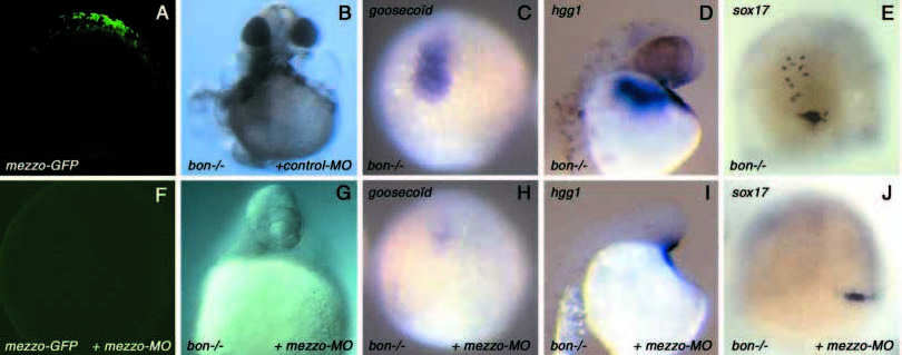
Fig. 8
Injection of an antisense morpholino oligonucleotide directed against mezzo in bon mutants. (A) Control embryo injected with 100 pg of an mRNA encoding a fusion protein between the 5′ leader sequence of mezzo and the sequence coding for the first 43 amino acids fused to the coding sequence of the green fluorescent protein to make mezzo-GFP mRNA. (F) Embryo injected with 100 pg of mezzo-GFP mRNA and with 12 ng the antisense morpholino directed towards the 5′ end of mezzo (mezzo-MO). All the embryos injected with mezzo-GFP mRNA were brightly fluorescent when examined at the shield stage (A). In embryos co-injected with mezzo-GFP and 12 ng of mezzo-MO, the fluorescence was abolished (F). Note that the camera gain in this image was much higher than for the image in A. Using the same settings as A, the image would be black. (B) bon mutants injected with an unrelated morpholino directed against the sea urchin hatching enzyme mRNA used as a negative control. (G) bon mutant embryo injected with 12 ng of antisense morpholino targeted against mezzo. (C,H) Expression of the prechordal plate marker goosecoid in control uninjected (C) and in morpholino injected (H) bon mutant embryos at 80% epiboly. (D,I) Expression of the hatching gland marker hgg1 in control uninjected (D) and in morpholino injected (I) bon mutants. (E,J) Residual sox17 expression in homozygous bon mutants at 80% epiboly (E), and absence of sox17-expressing cells in bon mutants injected with the morpholino (J). To unambiguously identify homozygous bon mutants, their genotype was determined by PCR (Kikuchi et al., 2001).