
|
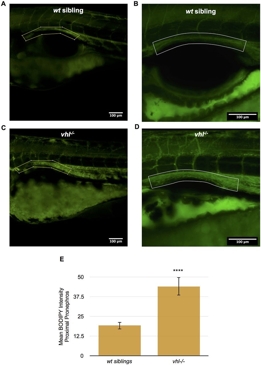
Fig. 3
Vhl-/- larvae have an increased amount of lipids in the proximal pronephros compared to wt siblings. (A-D) BODIPY 493/503 immunofluorescence was performed in live whole-mount 5.5dpf vhl-/- and wt siblings. BODIPY 493/503 staining (in green) intensity was then quantified within a ROI encompassing the proximal pronephros (yellow box), using ImageJ. Confocal images of BODIPY staining in vhl-/- (C) versus wt sibling (A). Confocal images of BODIPY staining in vhl-/- (D) versus wt sibling (B). (E) Quantification of mean BODIPY 493/503 staining intensity within the ROI in 5.5dpf vhl-/- versus wt siblings. Each experiment had at least three larvae per sample group and was performed in triplicate. Data represent mean±s.e.m. ****P<0.0001 (paired two-tailed t-test).