
Image
|
Figure Caption
Fig. S5
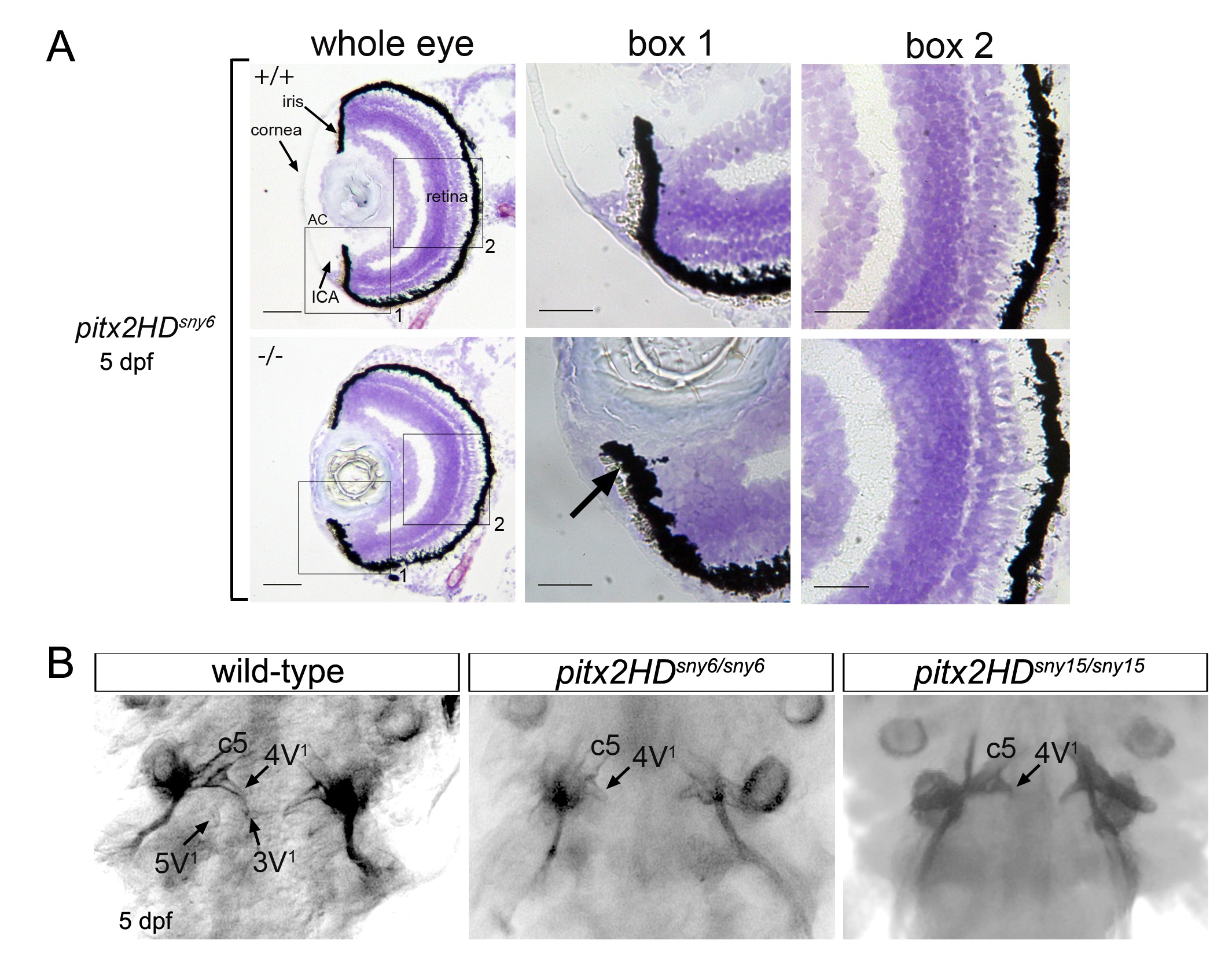
Pitx2HD mutants develop eye and tooth defects associated with ARS. (A) Cryosections of the eye from 5 dpf wild-type (+/+) or pitx2HDsny6/sny6 (-/-) fish stained with crystal violet. AC=anterior chamber; ICA=iridocorneal angle. Scale bars=50 µm. Box1 and Box2 are zoom-in views of boxed structures. Retina organization appeared similar in wild-type and pitx2HD mutant fish. (B) Ventral views of alizarin red staining of teeth at 5 dpf wild-type, pitx2HDsny6/sny6 and pitx2HDsny15/sny15 fish.
Figure Data
Acknowledgments
This image is the copyrighted work of the attributed author or publisher, and
ZFIN has permission only to display this image to its users.
Additional permissions should be obtained from the applicable author or publisher of the image.
Reprinted from Developmental Biology, 416(1), Ji, Y., Buel, S.M., Amack, J.D., Mutations in zebrafish pitx2 model congenital malformations in Axenfeld-Rieger syndrome but do not disrupt left-right placement of visceral organs, 69-81, Copyright (2016) with permission from Elsevier. Full text @ Dev. Biol.