
IMAGE
Fig. 2
- ID
- ZDB-IMAGE-160803-50
- Publication
- Quintana et al., 2014 - Hcfc1b, a zebrafish ortholog of HCFC1, regulates craniofacial development by modulating MMACHC expression
- All Figures
- Figures for Quintana et al., 2014
Image
|
Figure Caption
Fig. 2
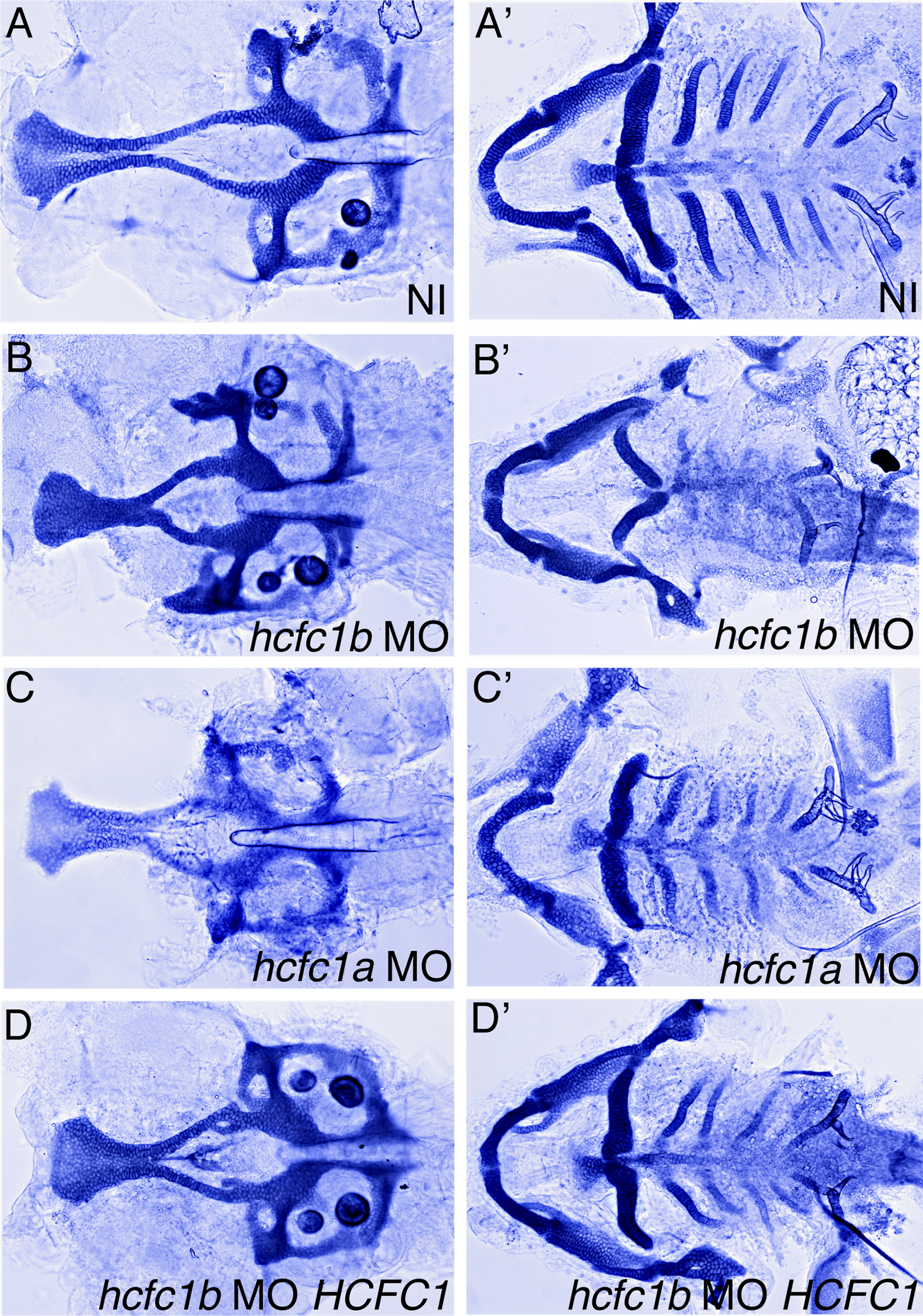
Loss of hcfc1b causes defects in craniofacial development. (A-D) Alcian:Alizarin staining was performed to visualize the developing cartilage in non-injected controls (NI), hcfc1a morphants (hcfc1a MO), hcfc1b morphants (hcfc1b MO), or embryos co-injected with hcfc1b MO and in vitro synthesized human HCFC1 mRNA. Embryos were stained at 5 days post fertilization and manual dissection of the viscerocranium and neurocranium was performed. Neurocranium is depicted in A-D and the viscerocranium is depicted in A′-D′.
Figure Data
Acknowledgments
This image is the copyrighted work of the attributed author or publisher, and
ZFIN has permission only to display this image to its users.
Additional permissions should be obtained from the applicable author or publisher of the image.
Reprinted from Developmental Biology, 396(1), Quintana, A.M., Geiger, E.A., Achilly, N., Rosenblatt, D.S., Maclean, K.N., Stabler, S.P., Artinger, K.B., Appel, B., Shaikh, T.H., Hcfc1b, a zebrafish ortholog of HCFC1, regulates craniofacial development by modulating MMACHC expression, 94-106, Copyright (2014) with permission from Elsevier. Full text @ Dev. Biol.