IMAGE

Fig. 2

ID
ZDB-IMAGE-160729-39
Genes
Source
Figures for Lai et al., 2014
Image
Figure Caption

Fig. 2

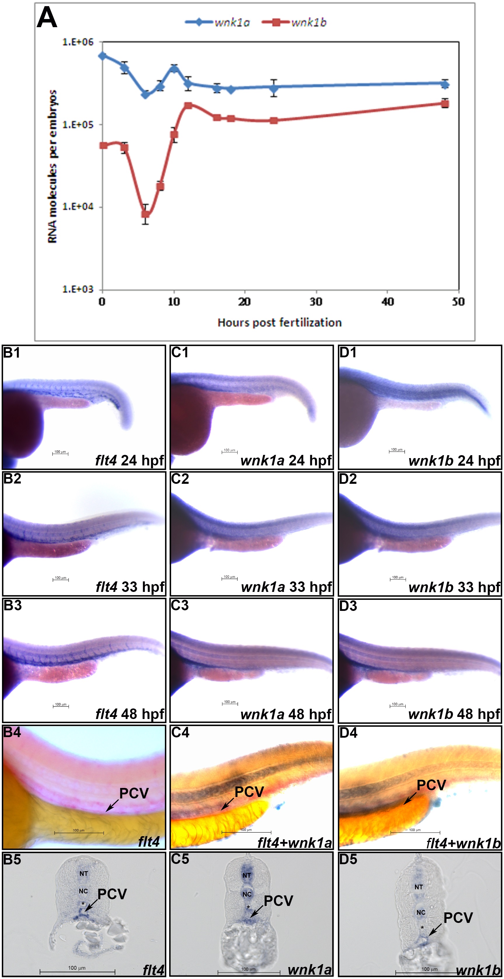
Spatial and temporal expression patterns of wnk1a and wnk1b.

(A) wnk1a and wnk1b mRNA expression profiles as determined by q-RT-PCR. At least three replicates were performed, and the average number of molecules was calculated using a standard curve from a q-RT-PCR assay. The standard deviations are shown in the graph. The red and blue lines indicate the wnk1a and wnk1b expression profiles, respectively. (Bin situ hybridization for flt4 (B) wnk1a (C), and wnk1b (D) was performed at the indicated time points. flt4 (B1wnk1a (C1~C3), and wnk1b (D1in situ hybridization for flt4 alone (B4), wnk1a+flt4 (C4), and wnk1b+flt4 (D4) shows the co-localization of wnk1a and wnk1b with flt4 in the PCV. Expression of flt4 (B5), wnk1a (C5) and wnk1b (D5) is seen in the PCV (arrow) in sections from stained embryos. wnk1a and wnk1b are also expressed in the neural tube (NT) and notochord (NC). The dorsal aorta (*) is negative for wnk1a and wnk1b expression. Scale bar: 100 µm.

Figure Data
Acknowledgments
This image is the copyrighted work of the attributed author or publisher, and ZFIN has permission only to display this image to its users. Additional permissions should be obtained from the applicable author or publisher of the image. Full text @ PLoS One