
|
Fig. 2
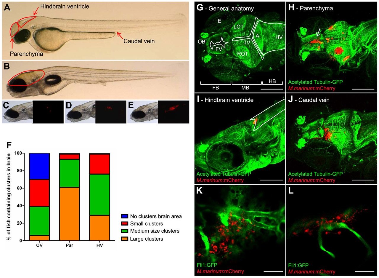
Three infection routes compared in the embryo model. (A) Photograph of a casper embryo at 2 dpf. Red arrows indicate the three different infection routes used. (B) Casper embryo at 7 dpf. The red line indicates the brain area. (C) Example of a small cluster. Left panel shows bright-field image; right panel shows corresponding fluorescent image. (D) Example of a medium cluster. (E) Example of a large cluster. (F) At 5 dpi, the infection was analyzed with fluorescence microscopy. Fluorescent bacterial clusters in the brain were counted visually. The clusters were scored, depending on their size, as small, medium or large. CV, caudal vein; Par, parenchyma; HV, hindbrain ventricle. (G–J) Z-stack of zebrafish embryos, at 7 dpf, stained with anti-acetylated tubulin (labelling axons; green) and infected with M. marinum E11 (red). Scale bars: 150 µm. (G) Anatomy of the zebrafish brain: FB, forebrain; MB, midbrain; HB, hindbrain; OT, optic tectum (L, left; R, right); OB, olfactory bulb; FV, forebrain ventricle; TV, tectal ventricle; HV, hindbrain ventricle; A, aquaductus; E, eye. (H) Embryo with multiple clusters after infection into the parenchyma. Arrow indicates a cluster in the ventricular wall. (I) Embryo infected into the hindbrain ventricle, with a cluster in the ventricle. Marked area indicates the hindbrain ventricle. (J) Embryo infected via the caudal vein, with a cluster in the parenchyma. (K,L) Tg(Fli1:GFP)y1 casper embryo (Fli1:GFP in green) at 5 days after infection via the caudal vein with M. marinum E11 (red). Both panels are single Z-slices and show the relationship between mycobacteria and blood vessels in the brain, which indicates migration of mycobacteria from bloodstream to brain tissue. Scale bars: 35 µm.