
|
Fig. 1
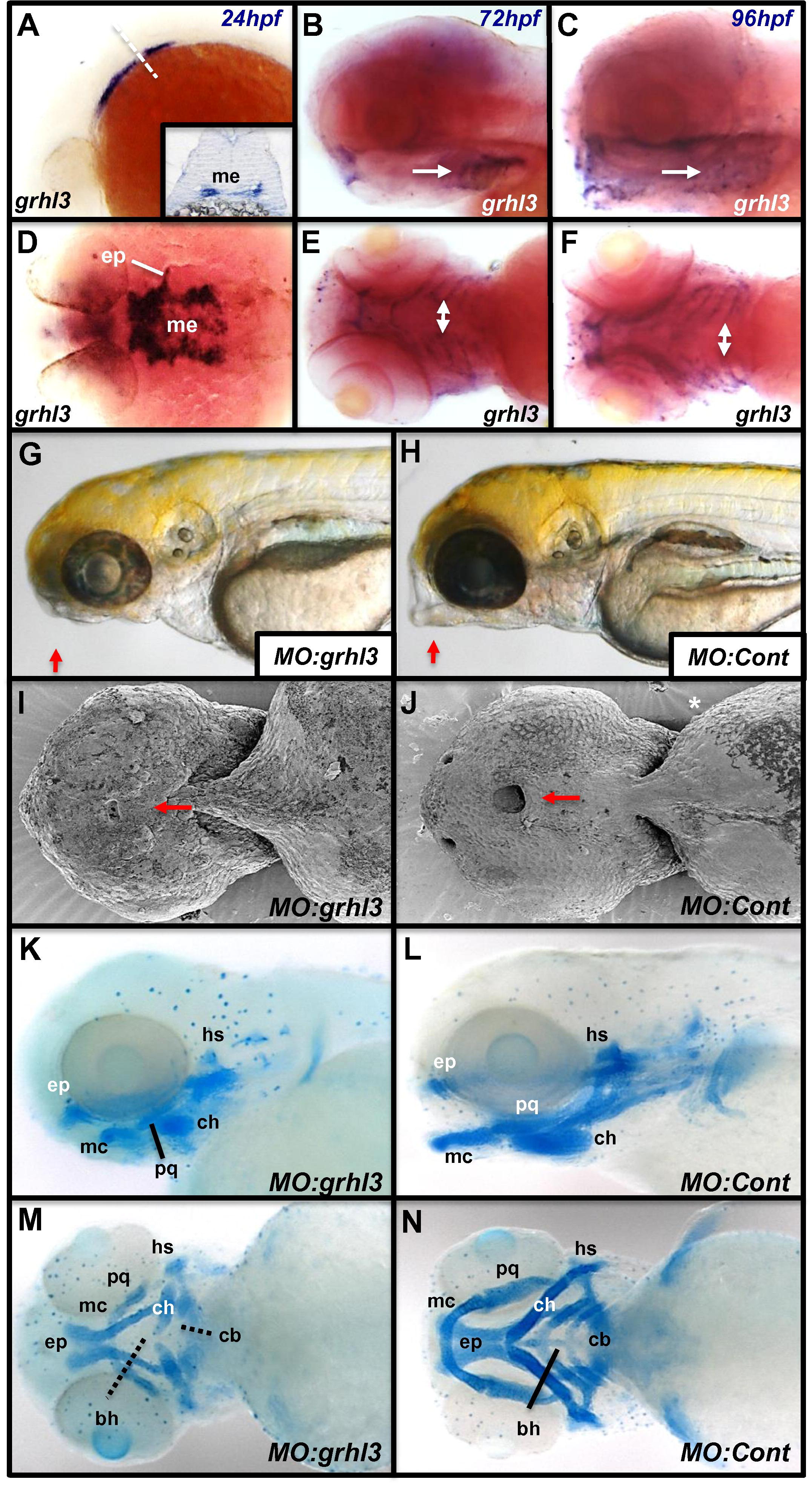
Loss of grhl3 causes defects in lower-jaw growth and development. (A-F) grhl3 is expressed in the medial endoderm (me) and developing endodermal pouches (ep) of the pharynx. Expression persists throughout the pharyngeal arches (white arrows) until at least 96 hpf (A-C, lateral view, anterior to left, inset in A shows cross-section in the plane of dotted line; D, dorsal view; E and F, ventral views). (G and H) Following MO-mediated knockdown of grhl3, fish exhibit defective craniofacial development when visualised by brightfield microscopy. (I and J) Scanning Electron Microscopy (SEM) at 48 hpf shows defective formation of the ventral region of the mouth (red arrows) in grhl3-morphants (I) relative to controls (J). (K–N) Lateral (K, L) and ventral (M, N) views of Alcian Blue stained cartilage displaying severe hypoplasia (palatoquadrate [pq], ceratohyal [ch] Meckel’s cartilage [mc]) or absence (basihyal [bh], ceratobranchials [cb]) of jaw structures in grhl3 morphants relative to controls. The hyosymplectic (hs) and ethmoid plate (ep) display minimal changes in grhl3 morphants at 96 hpf.
Reprinted from Mechanisms of Development, 133, Dworkin, S., Simkin, J., Darido, C., Partridge, D.D., Georgy, S.R., Caddy, J., Wilanowski, T., Lieschke, G.J., Doggett, K., Heath, J.K., Jane, S.M., Grainyhead-like 3 regulation of endothelin-1 in the pharyngeal endoderm is critical for growth and development of the craniofacial skeleton, 77-90, Copyright (2014) with permission from Elsevier. Full text @ Mech. Dev.