
|
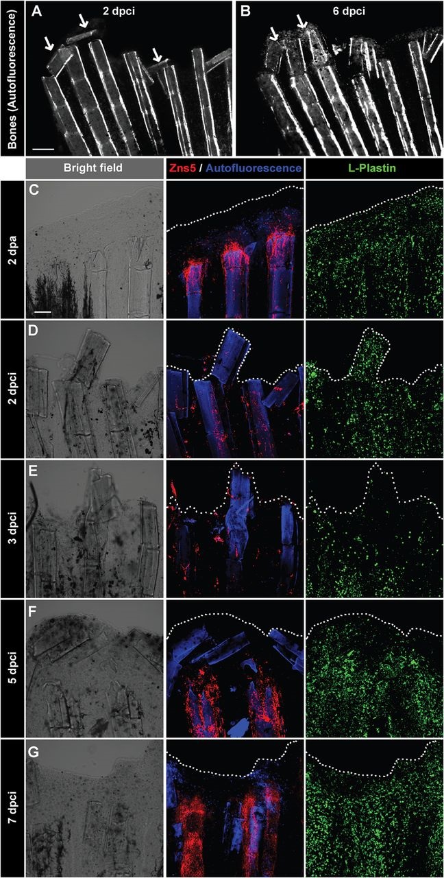
Fig. 5
Detachment of the destroyed fin tissue is associated with displacement and resorption of the dead bone fragments at the wound margin. (A,B) Imaging of bones in the same fin detected by autofluorescence of the mineralized matrix at 2 and 6dpci. The margin of the remnant fins contains detached and displaced bone fragments between the rays that become resolved (arrows). N=5. (C-G) Confocal imaging of whole-mount fins immunostained with the osteoblast marker Zns5 (red), phagocyte marker L-plastin (green) and autofluorescent bone matrix (blue) at 2dpa (C) and at different time points after cryoinjury (D-G). At 2dpa (C), Zns5-labelled osteoblasts accumulate at the tip of the bone to initiate bone regeneration. L-plastin-expressing cells are present in the entire tissue. At 2dpci (D) and 3dpci (E), osteoblasts are scattered along the bones in irregular manner. At 5dpci (F), Zns5-positive cells are enriched at the tips of the intact bones, below the margin of the stump that contains bone debris devoid of osteoblasts. At 7dpci (G), Zns5 immunostaining is robustly enhanced along the remaining bones, indicating resumed regeneration. L-plastin-expressing cells are associated with the repairing and regenerating tissue. N=4. Scale bar in A=200µm, in C=100µm.