
Image
|
Figure Caption
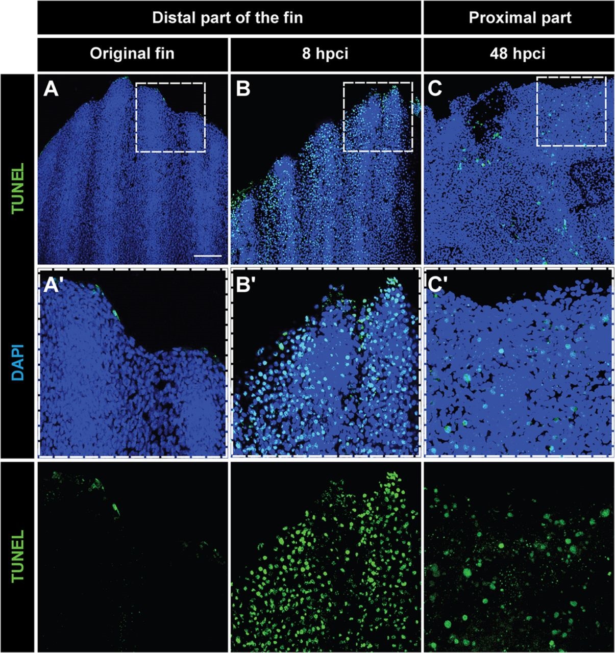
Fig. 2
Tissue loss after cryoinjury is associated with massive apoptosis. (A-C) Whole-mount staining with DAPI (blue) and TUNEL (green) of the original fin (A), at 8hpci (B) and at 48hpci (C). (A′-C′) Magnifications of the framed areas of the upper images. (A,A′) Uninjured fins contain few apoptotic cells at the distal margin. (B,B2) Before sloughing of the cryoinjured fin part at 8hpci, extensive apoptosis in the distal part of the extremity is observed. (C,C′) After truncation of the damaged fin part at 48hpci, the margin of the remaining stump still contains apoptotic cells. N=4. Scale bar in A=100µm.
Acknowledgments
This image is the copyrighted work of the attributed author or publisher, and
ZFIN has permission only to display this image to its users.
Additional permissions should be obtained from the applicable author or publisher of the image.
Full text @ Biol. Open