
|
Fig. S1
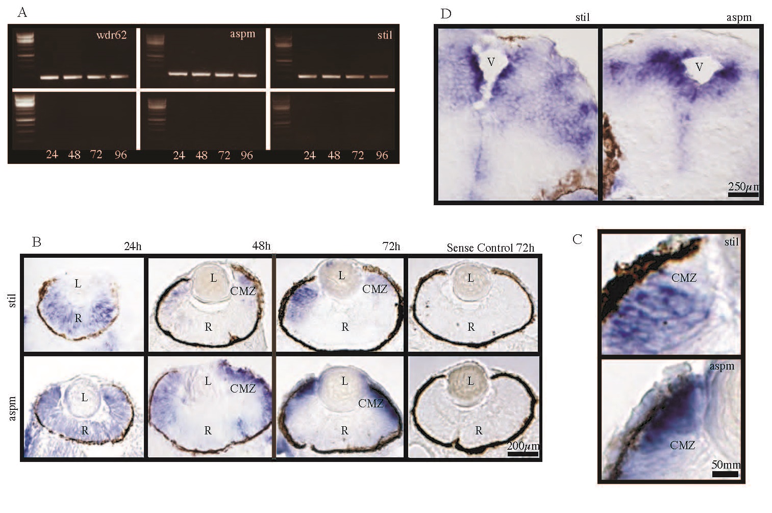
The MCPH genes stil, aspm and wdr62 are expressed in developing zebrafish embryos at early time points including 24hpf, 48hpf, 72hpf and 96hpf. In-situ hybridization (ISH) for stil and aspm demonstrated expression in the retina and brain, with localization to regions of active proliferation. (A) RT-PCR demonstrates expression of wdr62, aspm and stil at 24hpf, 48hpf, 72hpf and 96hpf alongside a 1kb DNA ladder. Bands correspond to predicted product sizes - 192bp (wdr62), 314bp (aspm) and 309bp (stil). Control PCR reactions (minus the RT step) are shown for each time point and condition, directly below each corresponding gel. (B) ISH of zebrafish retinal sections, using DIG-labelled antisense probes, demonstrates expression patterns of stil and aspm at 24hpf, 48hpf and 72hpf. Expression of stil is seen throughout the neural retina at 24hpf but by 48hpf expression occurs almost exclusively at the ciliary marginal zone (CMZ). Continued CMZ-specific expression of stil can be seen at 72hpf. Similarly, aspm is expressed throughout the neural retina at 24hpf. However, at 48hpf expression occurs mainly at the CMZ and in adjacent retinal tissue and by 72hpf strong aspm expression is seen in the CMZ, with weak to no staining elsewhere in the retina. Sense probe hybridization was performed as a control (shown here for each gene at 72hpf). (C) CMZ staining at 72hpf for both stil and aspm shown in high magnification. (D) ISH of zebrafish embryo brain sections at 72hpf demonstrates stil and aspm expression in the periventricular regions, where progenitor cells continue undergoing mitotic cell division.