
|
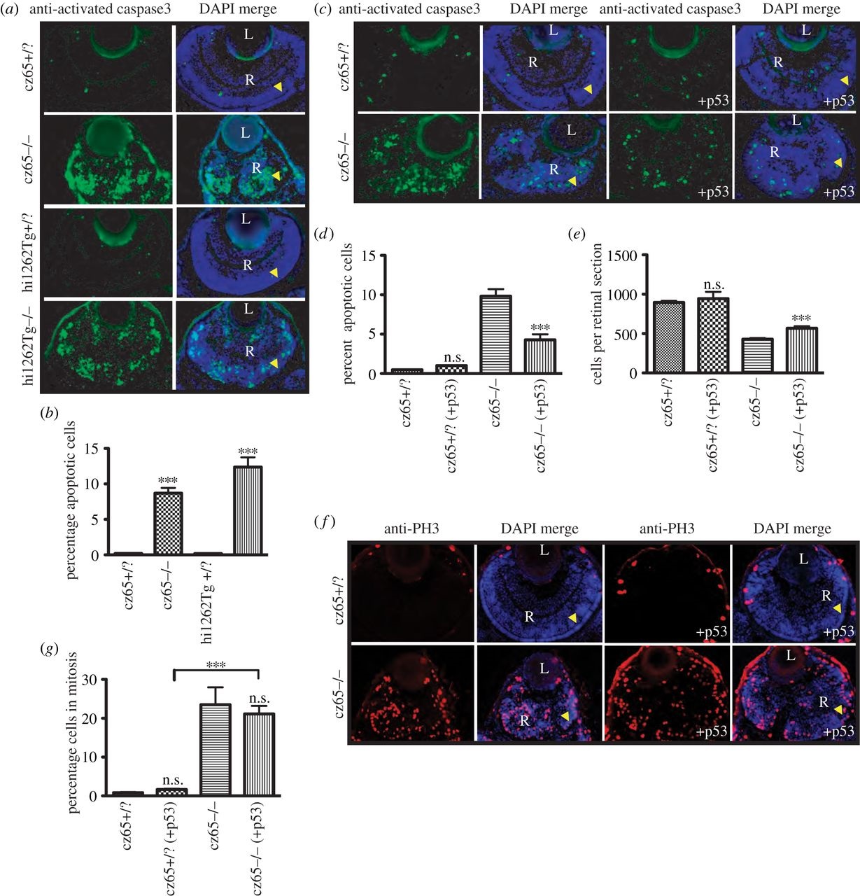
Fig. 6
Apoptosis is associated with the MCPH phenotype. (a,b) High levels of apoptotic cell death occur within the retina of developing stil mutant embryos. (a) Little or no apoptotic cell death, as marked by antiactivated caspase-3 antibody (green) was seen in stilcz65+/? retinas at 72 hpf (also shown with DAPI-counterstain in blue). By contrast, high levels of apoptotic cell death were seen throughout the retina of stilcz65-/- embryos. A similar pattern was seen in stilhi1262Tg-/- embryos, with little apoptotic death in stilhi1262+/? retinas, but high levels of apoptosis in the stilhi1262Tg-/- mutant. (b). In stilhi1262Tg-/- mutants at 72 hpf, 8.7% of retinal cells were observed to be undergoing apoptosis (n = 52) versus 0.2% in stilcz65+/? (n = 24) (p < 0.001). In stilhi1262Tg-/- embryos, 12.4% of retinal cells were apoptotic (n = 16) versus 0.2% in stilhi1262+/? (n = 22) (p < 0.001). (c-g) Blocking apoptosis in stil mutant embryos partially rescued the retinal phenotype but did not rescue the mitotic phenotype. (c-d) anti-p53 Mo injection led to a reduction in apoptosis (green; antiactivated caspase-3) in stilcz65-/- mutants at 72 hpf, from 9.8% of cells (n = 39) to 4.3% (n = 15), p < 0.001. By comparison, 0.47% of retinal cells underwent apoptosis in stilcz65+/? embryos (n = 38) with no significant difference with anti-p53 Mo (1.0%; n = 2), p > 0.05. This anti-p53 Mo-related reduction in apoptosis led to partial rescue of retinal size (c,e), with an increase in mean cells per retinal section to 568 (n = 15) from 429 (n = 39), p < 0.01. A smaller, non-significant increase in retinal cells was seen in stilcz65+/? embryos; 944 (n = 2) versus 895 (n = 38), p > 0.05). (f) While anti-p53 Mo reduces apoptosis and led to an increase in retinal size and cell number, there was no significant effect on the mitotic phenotype, as demonstrated by anti-PH3 immunostaining (red). (g) anti-p53 Mo injection had no significant effect on MI (stilcz65-/- with p53 Mo; 21.2% (n = 12) versus 23.5% (n = 7) for stilcz65-/- without p53 Mo, p > 0.5. As a control, anti-p53 Mo was also injected into stilcz65+/? embryos with no significant effect on percentage of mitotic cells (stilcz65+/? with p53 Mo 1.7% (n = 13) versus 0.8% (n = 3) in stilcz65+/? embryos without p53 Mo; p < 0.05. n = number of eyes analysed.