
Fig. 6
- ID
- ZDB-IMAGE-160707-25
- Publication
- Lisse et al., 2016 - Paclitaxel-induced epithelial damage and ectopic MMP-13 expression promotes neurotoxicity in zebrafish
- All Figures
- Figures for Lisse et al., 2016
|
Fig. 6
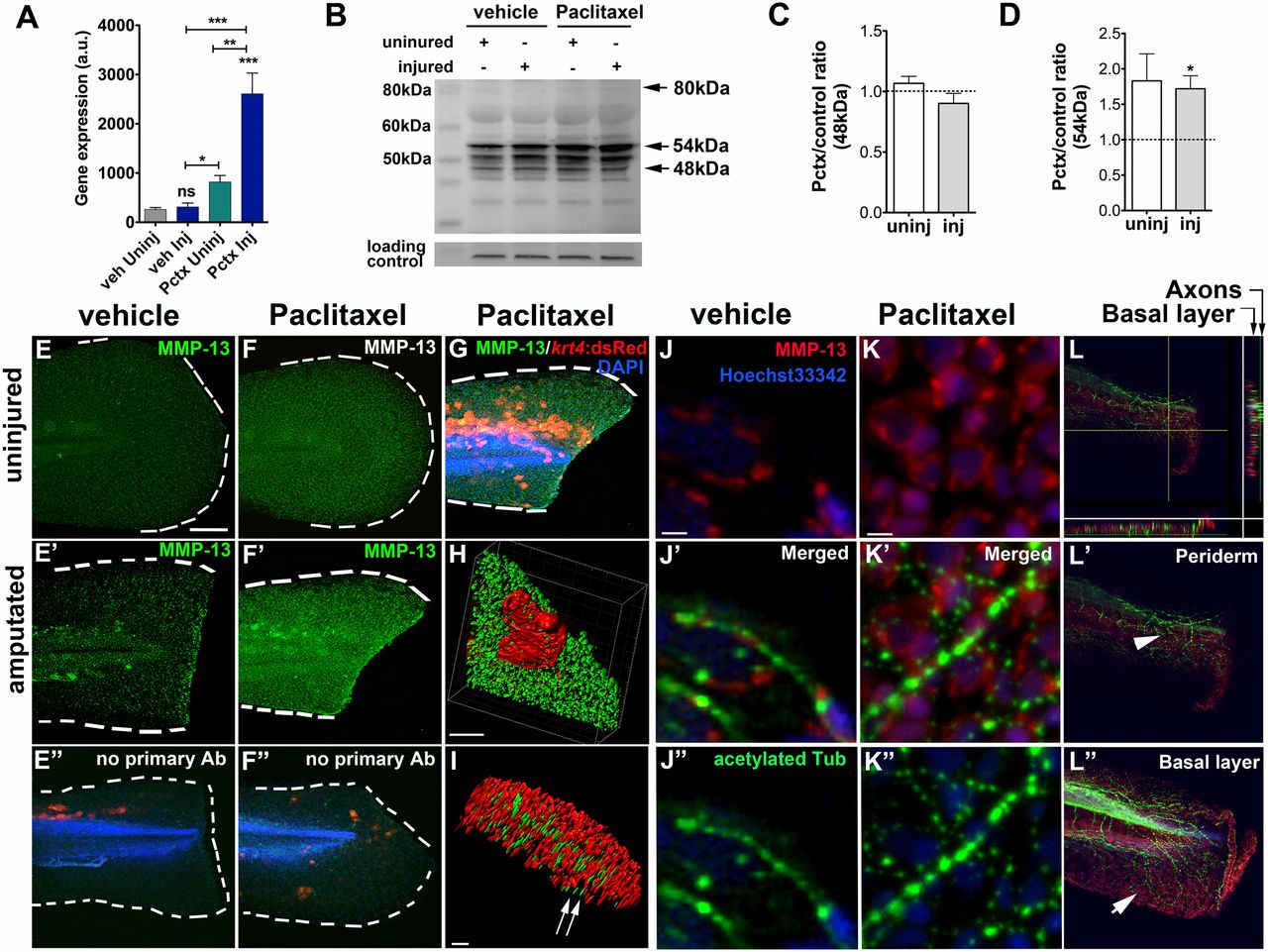
Paclitaxel stimulates MMP-13 expression. (A) Quantitative real-time PCR shows increased mmp13a expression in uninjured and injured animals treated with 22 µM paclitaxel for 3 h (15 pooled larvae per group). (B) Western analysis shows higher abundance of the 54-kDa isoform in uninjured and injured animals treated with paclitaxel for 3 h (10 pooled larvae per group). (C and D) Paclitaxel/vehicle ratios for normalized 48-kDa (C) and 54-kDa (D) bands in uninjured and injured animals (n = 2, 10 pooled larvae). Dashed lines demarcate control levels. (E-F′′) Immunofluorescence staining of MMP-13 in vehicle control (E) is increased at the wound margin after amputation (E′) and is ubiquitous following 3 h of paclitaxel treatment (F and F′). Immunofluorescence staining of larvae transiently injected with krt4:dsRed in the absence of primary MMP-13 antibody (E′′ and F′′). (Scale bar, 50 µm.) (G) Mosaic keratinocyte-specific expression (red) following krt4:dsRed injection and MMP-13 (green) staining shows colocalization. DAPI-stained nuclei. (Scale bar, 50 µm.) (H) 3D rendering of one keratinocyte (red) and MMP-13 staining (green) shows colocalization. (Scale bar, 10 µm.) (I) 3D rendering of axons (acetylated-tubulin, green) and MMP-13 staining (red) shows no colocalization (arrows). (Scale bar, 15 µm.) (J-K′′) MMP-13 staining (J and K) and axons stained with acetylated tubulin (J′′ and K′′) show lack of colocalization in vehicle (J′) and paclitaxel- (K′) incubated larvae. (Scale bars, 5 µm.) (L) Orthogonal views (sidebars) show axons (green) colocalizing with basal cell-specific MMP-13 staining (red). (L′ and L′′) MMP-13 staining (arrow in L′′) is present in the deeper basal layer below the axons (arrowhead) and is absent from the superficial periderm (L′). (Scale bar, 50 µm.) *P < 0.05, **P < 0.01, ***P < 0.001. AB, antibody; a.u., arbitrary units; Inj, injured; Pctx, paclitaxel; Uninj, uninjured; veh, vehicle.