
Image
|
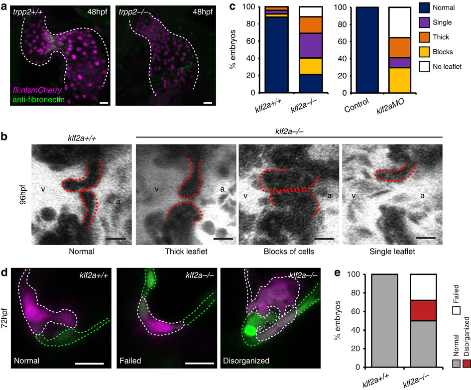
Figure Caption
Fig. 6
Characterization of klf2a-/- mutants.
(a) anti-fibronectin staining (green) is reduced in the AVC of trpp2-/- mutants (12/12 trpp2+/+, 9/19 trpp2-/-). (b,c) A range of valvular defects were observed in klf2a-/- mutant embryos at 96 hpf (n=25 klf2a+/+, n=48 klf2a-/-), similar to those observed in klf2aMO-injected embryos (e; n=20 control, n=17 klf2aMO). (d) Photoconversion of AVC EdCs at 48 hpf shows the organization of cells within the cardiac jelly to be affected by loss of klf2a (d,e; n=21 klf2+/+, n=32 klf2a-/-) at 72 hpf. Scale bars, 10 µm.
Figure Data
Acknowledgments
This image is the copyrighted work of the attributed author or publisher, and
ZFIN has permission only to display this image to its users.
Additional permissions should be obtained from the applicable author or publisher of the image.
Full text @ Nat. Commun.